Visualizing Vascular Volumes

Cerebral vascular images obtained through angiography are used by neurosurgeons for diagnosis, surgical planning and intra-operative guidance. The intricate branching of the vessels and furcations, however, make the task of understanding the spatial three-dimensional layout of these mages challenging. In this paper, we present empirical studies on the effect of different perceptual cues (fog, pseudo-chromadepth, kinetic depth, and depicting edges) both individually and in combination on the depth perception of cerebral vascular volumes and compare these to the cue of stereopsis. Two experiments with novices and one experiment with experts were performed. The results with novices showed that the pseudo-chromadepth and fog cues were stronger cues than that of stereopsis. Furthermore, the addition of the stereopsis cue to the other cues did not improve relative depth perception in cerebral vascular volumes. In contrast to novices, the experts also performed well with the edge cue. In terms of both novice and expert subjects, pseudo-chromadepth and fog allow for the best relative depth perception, although experts unlike novices also performed well with the edge cue. By using such cues to improve depth perception of cerebral vasculature we may improve diagnosis, surgical planning, and intra-operative guidance.

Publications

  1.  Marta Kersten-Oertel, S. J. S. Chen, D. Louis Collins. An Evaluation of Depth Enhancing Perceptual Cues for Vascular Volume Visualization in Neurosurgery. IEEE Trans Vis Comput Graph. March, 2014.
  2.  M. Kersten-Oertel, S. J. S. Chen, D. L. Collins. “A Comparison of Depth Enhancing Perceptual Cues for Vessel Visualization in Neurosurgery.” CARS, June 27–30, 2012.

Normal Aging

adni_model_4d_v2

Over the past several years we have collaborated with several individuals to study normal aging. Using Alzheimer’s disease, amnesic mild cognitive impairment and healthy individuals we have demonstrated that interhemispheric coupling may be regarded as a flexible mechanism that can improve the brain’s ability to meet processing demands for high cognitive demand in normal aging and for low cognitive demand in AD [1].

In a recent study the links between cognitive ability and cortical tissue volume in old age were investigated. Evidence from this research warns against an exclusive reliance on the causal link between cognitive function and the cortical tissue in old age based on assumptions of the aging process. Preservation of cortical tissue thickness in old age is not a foundation for successful cognitive aging, but rather reflects a lifelong association [2].

Reference

[1] J. Ansado, D.L. Collins, S. Joubert, V.S. Fonov, O. Monchi, S.M. Brambati, F. Tomaiuolo, M. Petrides, S. Faure, Y. Joanette, Yves. “Interhemispheric coupling improves the brain’s ability to perform low cognitive demand tasks in Alzheimer’s disease and high cognitive demand tasks in normal aging”, Neuropsychology, Vol 27(4), Jul 2013, 464–480.

[2] Karama S, Bastin ME, Murray C, Royle NA, Penke L, Muñoz Maniega S, Gow AJ, Corley J, Valdés Hernández Mdel C, Lewis JD, Rousseau MÉ, Lepage C, Fonov V, Collins DL, Booth T, Rioux P, Sherif T, Adalat R, Starr JM, Evans AC, Wardlaw JM, Deary IJ. Childhood cognitive ability accounts for associations between cognitive ability and brain cortical thickness in old age. Mol Psychiatry. 2014 May;19(5):555–9.

Brain Shift

Since the introduction of the first intraoperative frameless stereotactic navigation device, image guided neurosurgery has become an essential tool for many neurosurgical procedures due to its ability to minimize surgical trauma by allowing for the precise localization of surgical targets. The integration of preoperative image information into a comprehensive patient-specific model enables surgeons to preoperatively evaluate the risks involved and define the most appropriate surgical strategy. Perhaps more importantly, such systems enable surgery of previously inoperable cases by helping to locate safe surgical corridors through IGNS-identified non-critical areas.

For intraoperative use, neuronavigation systems must relate the physical location of a patient with the preoperative models by means of a transformation that relates the two through a patient-to-image mapping. Throughout the intervention, hardware movement, an imperfect patient-image mapping, and movement of brain tissue during surgery invalidates the patient-to-image mapping. These sources of inaccuracy, collectivey described as ‘brain shift’, reduce the effectiveness of using preoperative patient specific models intraoperatively. Intraoperative imaging, such as MRI, has been shown to improve the accuracy of tumour resections through lengthened image guidance. However, such technology is extremely expensive, prolongs surgery, poses logistical challenges during awake surgeries, and is available in only a few centres worldwide. We have developed a neuronavigation platform (IBIS Neuronav) that integrates tissue deformation tracking during surgery based on tracked intraoperative ultrasound (iUS) that can accurately align all pre-operative data to the iUS to account for brain shift throughout a surgical intervention.


Reference:

[1] I. Gerard and D. L. Collins, “An Analysis of Tracking Error in Image Guided Neurosurgery”, Int. J. Computer Assisted Radiolgy and Surgery. 2015, Jan 4; 1–10 [Epub ahead of print].

[2] H. Rivaz, D.L. Collins, “Near real-time robust non-rigid registration of volumetric ultrasound images for neurosurgery”, Ultrasound in Medicine and Biology. 2015 Feb; 41(2): 574–587.

[3] H. Rivaz, S.J.S Chen, D.L. Collins, “Automatic Deformable MR-Ultrasound Registration for Image-Guided Neurosurgery”, IEEE Transactions on Medical Imaging. 2015 Feb; 34(2); 366–380.

[4] H. Rivas, Z. Karimaghaloo, D.L. Collins, “Nonrigid Registration of Ultrasound and MRI Using Contextual Conditioned Mutual Information”, IEEE Trans Med Imag. 2014 Mar;33(3):708–25.

[5] S. Beriault, A. Sadikot, F. Alsubaie, S. Drouin, D.L. Collins, G.B. Pike. “Neuronavigation using susceptibility-weighted venography: application to deep brain stimulation and comparison with gadolinium contrast”, Journal of Neurosurgery. 2014 Jul;121(1):131–41.

[6] L. Mercier, D Araujo, C Haegelen, RF Del Maestro, K Petrecca, DL Collins, “Registering pre- and post-resection 3D ultrasound for improved residual brain tumor localization”, Ultrasound in Medicine and Biology, 2013 Jan;39(1):16–29.

[7] M. Kersten-Oertel, P. Jannin, D.L. Collins, “The State of the Art in Mixed Reality Visualization in Image-Guided Surgery”, IEEE Transactions on Visualization and Computer Graphics. 2013 Mar;37(2):98–112.

[8] D. De Nigris, D. L. Collins, T. Arbel, “ Fast Rigid Registration of Pre-Operative Magnetic Resonance Images to Intra-Operative Ultrasound for Neurosurgery based on High Confidence Gradient Orientations”, 2013 July; 8(4): 649–661.


Augmented Reality in Neurovascular Surgery

In neurovascular surgery, and in particular surgery for arteriovenous malformations (AVMs), the surgeon must map pre-operative images of the patient to the patient on operating room (OR) table in order to understand the topology and locations of vessels below the visible surface. This type of spatial mapping is not trivial, is time consuming, and may be prone to error. Using augmented reality (AR) we can register the microscope/camera image to pre-operative patient data in order to aid the surgeon in understanding the topology, the location and type of vessel lying below the surface of the patient. This may reduce surgical time and increasing surgical precision. In this project as well as studying a mixed reality environment for neuromuscular surgery, we will examine and evaluate which visualization techniques provide the best spatial and depth understanding of the vessels beyond the visible surface.

A: Colour coding of a vascular DS-CTA volume based on blood flow. B: Vessels overlaid on the patient skin prior to draping (left). The AR view is used at this step to help tailor the extent of the craniotomy. On the right we see vessels overlaid on the cortex prior to resection, here the AR view is used to determine the optimal resection corridor. The blue arrows point to the pink markers that indicate the location of deep feeding arteries. The orange arrow indicates the major arterialized vein, shown as red and not blue. C: Different visualization techniques for combining the live camera image (prior to resection) with the virtual vessels (green, red, blue) are shown.The use of simple alpha-blending between the real and virtual worlds does not provide spatial information (top). More sophisticated techniques such as modulating transparency in the area of interest and using edges (from the virtual vessels and/or camera image) and using fog are applied. D: Based on the virtual information the surgeon placed a micropad on the brain surface above a virtual marker representing a deep feeding artery to help with the resection approach and vessel localization.

Publications

M. Kersten-Oertel, M., Gerard, I., Drouin, S., Mok, K., Sirhan, D., Sinclair, D. S. and Collins, D. L. Augmented reality in neurovascular surgery: feasibility and first uses in the operating room. IJCARS (2015): 1–14.

Kersten-Oertel, M., Gerard, I. J., Drouin, S., Mok, K., Sirhan, D., Sinclair, D. S., & Collins, D. L. (2015). Augmented Reality for Specific Neurovascular Surgical Tasks. In Augmented Environments for Computer-Assisted Interventions (pp. 92–103). Springer International Publishing.

M. Kersten-Oertel, I. Gerard, S. Drouin, K. Mok, D. Sirhan, D. Sinclair, D. L. Collins. “Augmented Reality in Neurovascular Surgery: First Experiences.” Augmented Environments for Computer-Assisted Interventions. Lecture Notes in Computer Science Volume 8678, 2014, pp 80–89, 2014.

Epilepsy

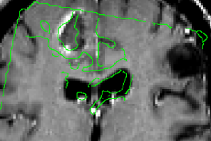
In some patients with refractory epilepsy that are candidates for surgery, intracranial EEG is recorded to precisely localize the epileptic focus. To record intracranial EEG, multiple depth electrodes are surgically implanted through holes in the skull, each with 8–15 equally spaced contacts. Current implantation planning consists of visual inspection of the patient’s MRI & CTA, visually searching for paths to the targets while avoiding vessels. This procedure is sub-optimal since estimation of the number of electrodes needed to sample a region and the precise location of each contact, which is paramount to accurately identify the focus and its extent, cannot be considered.

The goal of this project, conducted by Dr. Zelmann, one of my postdoctoral fellows, is to evaluate the clinical use of optimized depth electrode planning. We hypothesize that the use of our computed aided procedure will increase the accuracy and amount of information obtained during EEG intracranial investigation while constraining the trajectories to safe paths.

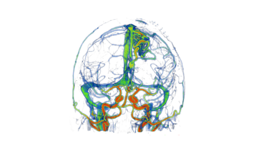
We have developed a procedure to optimize electrode location that would enable us to obtain complete coverage of a lesion or region of interest and surrounding neocortical grey matter, while minimizing the risk of approaching vessels and other critical structures. To this end, we automatically segment MRI, CT and CTA data; we model each electrode as a cylinder to assess risk; we estimate the contribution of individual contacts to record EEG; we compute an aggregated score for each electrode and a global score, obtaining the best cohort as the combined set of electrodes that remain at a safe distance.

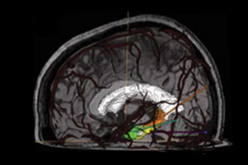
To allow its clinical use during planning and surgical implantation, the procedure is integrated into an image-guided neuronavigation system developed in our research laboratory over the past decade, which is called IBIS (Interactive Brain Imaging System; Mercier et al., Int J CARS. 2011;6(4):507–522). IBIS allows planning and navigation based on preoperative medical image data. The best cohort and a list of possible electrodes per target (ordered in terms of risk and recorded EEG) is generated and displayed in IBIS and will be available during planning and in the OR. The surgeon can visually review trajectories, decide between one of the automatic trajectories and the manual one and, if necessary, re-plan a trajectory in almost real time.

Preliminary results [1] presented at an international workshop on clinical image-based procedures, suggest that automatic planning allows recording from a larger volume than manually planned trajectories (p<0.01) and from more temporal grey matter (p<0.001), while remaining further away from segmented vessels (p<0.01). We are currently evaluating the procedure in retrospective clinically acquired imaging data from 20 patients with electrodes implanted in MTL regions. We are comparing visual and automatic trajectories by estimating volume recording from the target volumes (in this case: amygdala and hippocampus) and neocortical temporal grey matter as well as distance to vessels and other critical structures.

Reference

[1] Zelmann, R., Beriault, S., Mok, K., Haegelen, C., Hall, J., Pike, G. B., Olivier, A & Collins, D. L. (2014). Automatic Optimization of Depth Electrode Trajectory Planning. In Clinical Image-Based Procedures. Translational Research in Medical Imaging (pp. 99–107). Springer International Publishing.

[2] R. Zelmann, S. Beriault, K. Mok, C. Haegelen, J. Hall, G.B. Pike, A. Olivier & D.L. Collins, “Improving Recorded Volume in Mesial Temporal Lobe by Optimizing Stereotactic Intracranial Electrode Implantation Planning” submitted to International Journal of Computer Assisted Radiology and Surgery (CARS-D-14–00251), Oct 9, 2014.

[3] Zelmann, R., Beriault, S., Mok, K., Haegelen, C., Hall, J., Pike, G. B., Olivier, A & Collins, D. L. (2014). Automatic Optimization of Depth Electrode Trajectory Planning. In Clinical Image-Based Procedures. Translational Research in Medical Imaging (pp. 99–107). Springer International Publishing.

[4] R. Zelman, D.L. Collins, “Automatic Optimization of Depth Electrode Trajectory Planning”, MICCAI 2013 Workshop on Clinical Image-based Procedures: Translational Research in Medical Imaging

[5] C. Haegelen, P. Perucca, C.-E. Châtillon, L. Andrade-Valença, R. Zelmann, J. Jacobs, D. L. Collins, F. Dubeau, A. Olivier and J. Gotman. “High-frequency oscillations, extent of surgical resection and surgical outcome in drug-resistant focal epilepsy”, Epilepsia 2013 May;54(5):848–57.

[6] Zelmann, R., S. Beriault, M. M. Marinho, K. Mok, J. A. Hall, N. Guizard, C. Haegelen, A. Olivier, G. B. Pike, and D. L. Collins. “Improving recorded volume in mesial temporal lobe by optimizing stereotactic intracranial electrode implantation planning.” International journal of computer assisted radiology and surgery (2015): 1–17.

Multiple Sclerosis

Multiple sclerosis (MS) is a neurological disease that predominately affects young adults. Inflammatory mechanisms were believed to be the main contributor to the development of MS. However, more recent neuropathological and magnetic resonance imaging (MRI) studies suggest that neurodegenerative processes play an equivalent central role, that these degenerative processes commence during the early stages of the disease, and that neuronal and axonal loss may be the key substrate for the development of disability. One macroscopic hallmark of neurodegeneration is brain atrophy, which can be readily investigated non-invasively using MRI. We have recently demonstrated brain atrophy in pediatric-onset MS patients, further supporting a very early and possibly primary role for neurodegeneration in MS.

The three current hypotheses for the specific pathobiology underlying brain atrophy in MS are:

  • 1) atrophy in focal lesions and normal-appearing brain tissue occurs secondary to neuroaxonal injury and Wallerian degeneration and loss of normal myelin or reduction in myelin density associated with inflammation-mediated tissue insult;
  • 2) atrophy is due to a diffuse, primary degenerative process associated with neuronal cell death and subsequent loss of axons and myelinated pathways; and
  • 3) atrophy is due to a combination of mechanisms 1 and 2. In children with MS, insult to precursors of primary myelination may further impede normal brain maturation contributing to failure of age-expected brain growth in addition to atrophic loss of established neural networks.

The onset of MS during childhood and adolescence provides a potentially enhanced capacity to distinguish the earliest aspects of MS pathobiology, as the young age of such patients inherently limits the time period available for subclinical disease. A further unique aspect of pediatric onset MS is the potential deleterious impact of MS pathobiology to the processes of primary myelination and normal brain maturation, and thus potential consequence of MS contributing to failure of age-expected brain growth. We have recently reported reduced brain volumes in pediatric-onset MS compared to pediatric normal controls in cross-sectional studies. We also noted that the reduction in brain volume was not only global, but also specifically notable in the thalamus. We now propose to delineate whether the reduced brain volumes reflect age-expected failure of normative growth or loss of previously developed brain tissue (atrophy), or both, and will further explore the selective vulnerability of specific brain regions.

MRI confirmation of progressive brain volume loss, detectable in children and adolescents with MS, will not only refute the concept of pediatric brain resiliency and enhanced repair, but will also emphasize the fundamental nature of neurodegenerative biology of MS. Such confirmation has significant import on future therapeutic strategies, as it implies that anti-inflammatory therapies alone may fail to mitigate the negative impact of MS, and that neuroprotective strategies will be required from onset. This study is supported by an operating grant (Biomedical Research) provided by the Multiple Sclerosis Society of Canada & The Multiple Sclerosis Scientific Research Foundation.

Reference

[1] R. Harmouche, N.K. Subbanna, D.L. Collins, D.L. Arnold, T. Arbel, “Probabilistic multiple sclerosis lesion classification based on modeling regional intensity variability and local neighbourhood information”, IEEE Transactions on Biomedical Engineering, 2015 May; 62(5): 1281–92.

[2] N Guizard, P Coupé, VS Fonov, JV Manjón, DL Arnold, DL Collins, “Rotation-invariant multi-contrast non-local means for MS lesion segmentation (RMNMS)”, NeuroImage: Clinical. 2015 May 8: 376–89.

[3] K. Weier, B. Banwell, A. Cerasa, D.L. Collins, A. Dogonowski, H. Lassmann, A. Quattrone, M.A. Sahraian, H.R. Siebner and T. Sprenger, “The role of the cerebellum in multiple sclerosis”, Cerebellum. 2015 Jun; 14(3); 364–74

[4] Aubert-Broche B, Fonov VS, Garcia-Lorenzo D, Mouiha A, Guizard N, Coupé P, Eskildsen SF, Collins DL. A new method for structural volume analysis of longitudinal brain MRI data and its application in studying the growth trajectories of anatomical brain structures in childhood. Neuroimage 2013;82C:393–402.

[5] Aubert-Broche B, Fonov V, Ghassemi R, Arnold DL, Banwell B, Sled JG, Collins DL. Regional brain atrophy in children with multiple sclerosis. Neuroimage 2011;58:409–415.

[6] Kerbrat A, Aubert-Broche B, Fonov V, Narayanan S, Sled JG, Arnold DL, Banwell B, Collins DL. Reduced head and brain size for age and disproportionately smaller thalami in child-onset MS. Neurology 2012;78:194–201.

Parkinson’s Disease

Parkinson’s disease (PD) is the second most common neurodegenerative disease, after Alzheimer’s disease, worldwide. At the NIST Lab, we are dedicated to explore potential structural biomarkers for the disease, as well as proposing image processing techniques for the surgical treatment of PD.


Projects include:

Stereotaxic surgery for movement disorders

Stereotaxic surgery for movement disorders

We have recently developed techniques [1] used to create a lower resolution 3D atlas, based on the Schaltenbrand and Wahren print atlas, which was integrated into a stereotactic neurosurgery planning and visualization platform (VIPER), and a higher resolution 3D atlas derived from a single set of manually segmented histological slices containing nuclei of the basal ganglia, thalamus, basal forebrain, and medial temporal lobe. We have therefore developed, and are continuing to validate, a high-resolution computerized MRI-integrated 3D histological atlas, which is useful in functional neurosurgery, and for functional and anatomical studies of the human basal ganglia, thalamus, and basal forebrain.

Parkinson’s disease (PD) is a neurodegenerative disorder that impairs the motor functions. Deep brain stimulation (DBS) is an effective therapy to treat drug-resistant PD. Accurate placement of the DBS electrode deep in the brain under stereotaxic conditions is key to successful surgery [2]. Accuracy depends on a number of factors including registration error of the stereotaxic frame, geometric distortion of the MRI scans and brain tissue shift during resulting from cerebrospinal fluid (CSF) leakage, cranial pressure change, and gravity after the burr-hole is opened.

By scanning through acoustic skull windows, transcranial ultrasound can provide non-invasive visualization of internal brain structures (i.e. midbrain, blood vessels, and certain nuclei like the substantia nigra) as well as metallic surgical instruments (i.e. DBS electrode and cannula). We believe that such images can be used to improve stereotaxic accuracy.

In the past decade, we have developed a prototype image-guided neuronavigation system called IBIS (Interactive Brain Imaging System) in our research laboratory, which enables the acquisition of intraoperative 2D/3D ultrasound, and addresses the issue of registration errors caused by brain shift by using ultrasound data to improve the patient/image alignment. By linking the preoperative MRI, and the corresponding surgical plan, to the transcranial ultrasound with appropriate registration methods, we will enable real-time monitoring of the DBS implantation and will improve the safety and accuracy of the procedure. Our goal is to acquire transcranial ultrasound images, and examine its performance as an intraoperative imaging modality.

The study, as well as surgical treatment of PD necessitate the delineation of basal ganglia nuclei morphology. Few automatic volumetric segmentation methods have attempted to identify the key brainstem substructures including the subthalamic nucleus (STN), substantia nigra (SN), and red nucleus (RN) due to their small size and poor contrast in conventional MRI. I recently developed a technique [3] with my Ph.D. student Yimming Xiao based on a dual-contrast patch-based label fusion method to segment the SN, STN, and RN. Our proposed method outperformed the state-of-the-art single-contrast patch-based method for segmenting brainstem nuclei using a multi-contrast multi-echo FLASH MRI sequence. This method is encouraging as it will provide promising developments for the treatment and research of PD. This study is supported by the NSERC/CIHR Collaborative Health Research Program

Reference

[1] A. F. Sadikot, M. M. Chakravarty, G. Bertrand, V. V Rymar, F. Al-Subaie and D. L. Collins. “Creation of computerized 3D MRI-integrated atlases of the human basal ganglia and thalamus”, Frontiers in Systems Neuroscience, 2011;5:71

[2] Y. Xiao, V.S. Fonov, S. Beriault, F. Al Soubaie, M.M. Chakravarty, A.F. Sadikot, G.B. Pike and D.L. Collins, “Multi-contrast unbiased MRI atlas of a Parkinson’s disease population”, Int J Comput Assist Radiol Surg. 2015 March; 10(3):329–41.

[3] Xiao Y, Fonov VS, Beriault S, Gerard I, Sadikot AF, Pike GB, Collins DL. Patch-based label fusion segmentation of brainstem structures with dual-contrast MRI for Parkinson’s disease. Int J Comput Assist Radiol Surg. 2015 July; 10(7):1029–41

[4] M.M. Chakravarty, G. Bertrand, C. Hodge, A.F. Sadikot, and D.L. Collins, “The creation of a brain atlas for image guided neurosurgery using serial histological data,” NeuroImage. 2006; 30(2): 359–76.

Autism Spectrum Disorder

Autism Spectrum Disorder (ASD) is a set of neurological disorders characterized by impaired social communication and interaction, as well as restricted, repetitive patterns of behaviour, interests and activities. It is commonly diagnosed at around the ages of 3 to 4 years, developing tools that could aid in obtaining a diagnosis as early as 6 or 12 months of age would allow an early therapy that could potentially increase the adaptation to society of children with ASD.

At the NIST, we have utilized voxel-wise image processing methods to investigate the brain development of children at high-risk of ASD during the first 2 years of life. This work has led to observations of significant differences in the growth trajectories of several regions in the brain between children who are diagnosed with ASD at 2 years of age and normal controls.

YouTube-logo-full_color