In image-guided surgery the surgeon must map pre-operative patient images from the navigation system to the patient on operating room (OR) table in order to understand the topology and locations of the anatomy of interest below the visible surface. This type of spatial mapping is not trivial, is time consuming, and may be prone to error. Using augmented reality (AR) we can register the microscope/camera image to pre-operative patient data in order to aid the surgeon in understanding the topology, the location and type of vessel lying below the surface of the patient. This may reduce surgical time and increasing surgical precision.
HIV enters the brain soon after seroconversion and potentially causes cognitive impairment. Although the incidence of severe dementia has been reduced, perhaps due to effective HIV treatment, the prevalence of mild to moderate cognitive impairment appears to be increasing. It has been reported that 30-50% of HIV+ patients with well-controlled infections show cognitive deficits. Several factors are thought to contribute to this brain injury. However, the literature has yet to produce a clear consensus of the mechanisms that may underlie brain injury.
Brain volume loss associated with a history of severe HIV-related immunosuppression.
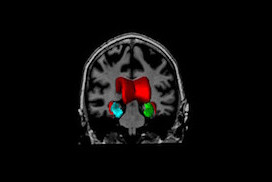
At NIST, we have utilized novel neuroimaging methods with complementary strengths, deformation-based morphometry, voxel-based morphometry and cortical modeling, to investigate the effects HIV has on brain structure and function. Here, we observed regionally specific patterns of reduced cortical and subcortical volumes in the HIV+ group. White matter loss and subcortical atrophy was related a history of more severe immunosuppression, while cortical thickness reductions were related to poorer neuropsychological test performance. The findings suggest that distinct mechanisms may underlie cortical and subcortical injury, and argues for the potential importance of early HIV treatment in protecting long term brain health.
Abstracts and Conference Presentations:
R. Sanford, A.L. Fernandez Cruz, L.K. Fellows, B.M. Ances, D.L. Collins, Regionally Specific Cortical Thinning in HIV+ Patients in the cART Era, 2016 Conference on Retroviruses and Opportunistic Infections (CROI), February 2016, Boston, Massachusetts (pdf)
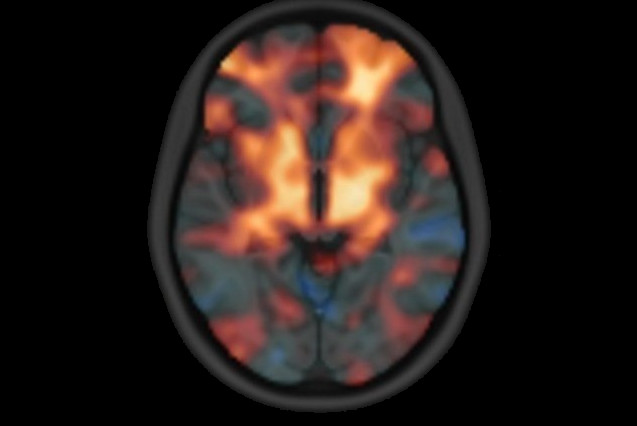
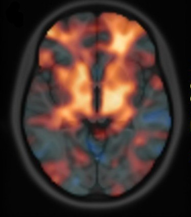
Alzheimer’s disease (AD) is a progressive neurodegenerative disease, which is the most common cause of dementia. Its prevalence for people aged 65-70 y is 1%, while it is 7% for the 75-84 y group, and 26% among those aged 90 and older. In Canada, persons over age 65 will make up to 15% of the population by 2016, but this amount is estimated to reach 23% by 2041. It is assumed that the prevalence of AD will quadruple by 2050 resulting in great financial burden.
It is believed that the pathophysiological process of AD begins well before the diagnosis of the dementia. Like many other neurodegenerative diseases, early treatment, before occurrence of too much irreversible degeneration of brain tissue, can be more effective. However, early diagnosis of Alzheimer’s disease is currently almost impossible. The well-cited biomarker model that the structural MRI begins showing abnormality at the preclinical stage and rises significantly in MCI stage, which makes it an interesting candidate for prognosis of dementia onset. Our group with sophisticated image analysis techniques followed by statistical analysis tries to make the diagnosis and prediction possible at the level of an individual person.
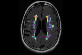
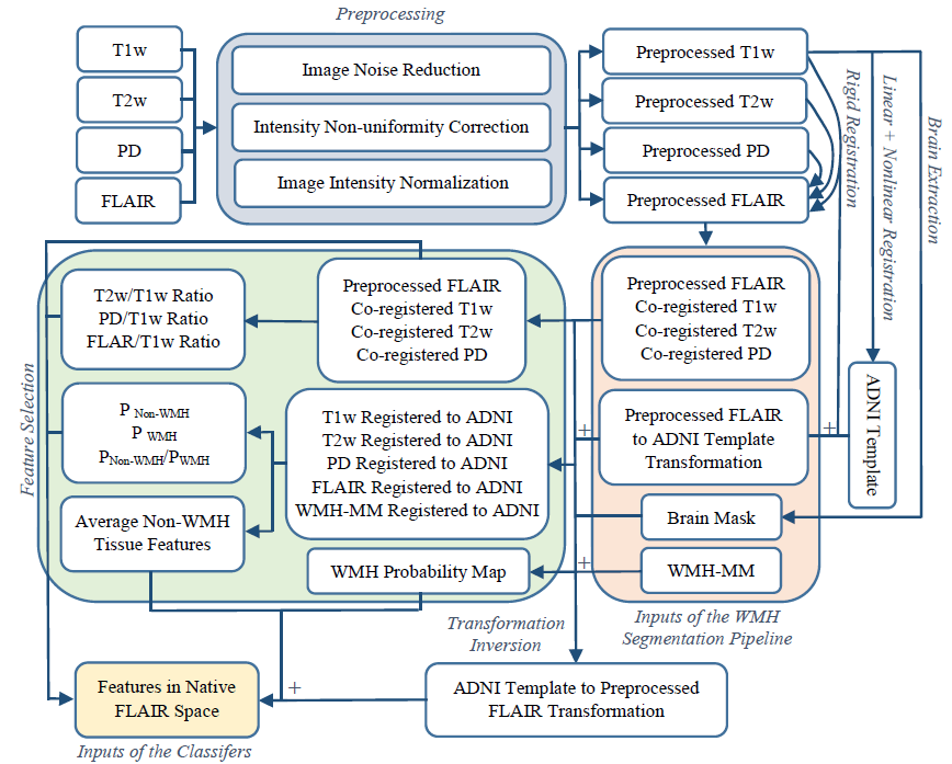
Neurodegenerative diseases such as Alzheimer’s disease (AD) commonly coexist with cerebrovascular disease in the elderly population. Cerebral small vessel disease (SVD) is the most common vascular cause of dementia and a major contributor to mixed dementia. SVD frequently coexists with AD and can increase the cognitive and physical deficits caused by neurodegeneration. White matter hyperintensities (WMHs) are considered to be one of the major signs of SVD on MRI and are associated with neurological and cognitive symptoms and physical difficulties.We have developed automated tools for segmentation of WMHs in Alzheimer’s patients using multiple contrasts of MR images. We use these segmentations to study the effect of WMHs in AD.
Dadar, M., Maranzano, J., Ducharme, S., Collins, D. L., & Alzheimer’s Disease Neuroimaging Initiative. (2019). “White Matter in Different Regions Evolve Differently During Progression to Dementia”. Neurobiology of Aging.
Dadar M., Zeighami Y., Yau Y., Fereshtehnejad S.M., Maranzano J., Postuma R.B., Dagher A., Collins D.L. (2018), “White Matter Hyperintensities Are Linked to Cognitive Decline in de Novo Parkinson’s Disease Patients”. NeuroImage: Clinical, 20, 892-900.
Dadar, M., Fonov, V. S., Collins, D. L., & Alzheimer’s Disease Neuroimaging Initiative. (2018). A comparison of publicly available linear MRI stereotaxic registration techniques. NeuroImage, 174, 191-200.
Dadar, M., Maranzano, J., Ducharme, S., Carmichael, O. T., Decarli, C., Collins, D. L., & Alzheimer’s Disease Neuroimaging Initiative. (2018). Validation of T 1w‐based segmentations of white matter hyperintensity volumes in large‐scale datasets of aging. Human brain mapping, 39(3), 1093-1107.
Dadar, M., Maranzano, J.,…, Collins, D.L. & Alzheimer’s Disease Neuroimaging Initiative. (2017). Performance comparison of 10 different classification techniques in segmenting white matter hyperintensities in aging. NeuroImage, 157, 233-249.
Dadar, M., Pascoal, T. A., Manitsirikul, S., Misquitta, K., … & Collins, D. L. (2017). Validation of a regression technique for segmentation of white matter hyperintensities in Alzheimer’s disease. IEEE transactions on medical imaging, 99, 1-1.