Augmented Reality Vessel Visualization

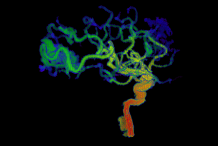
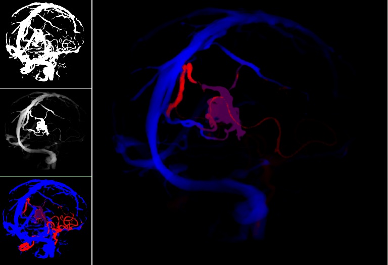
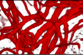
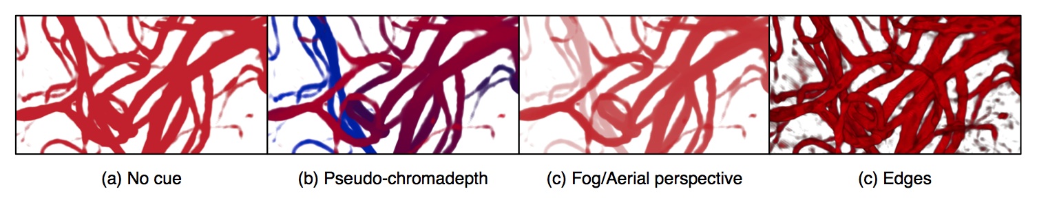
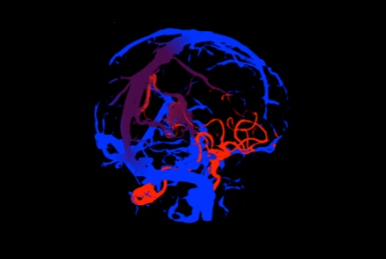
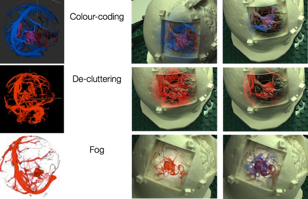
We explored a number of different volume rendering methods for AR visualization of vessel topology and blood flow.

Publications

  1. M. Kersten-Oertel, S. Drouin, S. J. S. Chen, D. L. Collins. ‘ “Volume Visualization for Neurovascular Augmented Reality Surgery”. Augmented Reality Environments for Medical Imaging and Computer-Assisted Interventions. Lecture Notes in Computer Science Volume 8090, pp 211–220, 2013.
  2. M. Kersten-Oertel, S.J.S Chen, S. Drouin, D. Sinclair, D. L. Collins. “Augmented Reality Visualization for Guidance in Neurovascular Surgery.” Stud Health Technol Inform 173:225–9. Proceedings of Medicine Meets Virtual Reality (MMVR), New Port, CA, Feb 9–11, 2012.
  3. S. Drouin, M. Kersten-Oertel, S. J. S. Chen, D. L. Collins. “A Realistic Test and Development Environmentfor Mixed Reality in Neurosurgery”. Augmented Environments for Computer Assisted Interventions’ (Proceedings of MICCAI AE-CAI Workshop 2011) Lecture Notes in Computer Science, Volume 7264:13–23, 2012.

Visualizing Blood Flow

Cerebral arteriovenous malformations (AVMs) are a type of vascular anomaly consisting of large intertwined vascular growth (the nidus) that are prone to serious hemorrhaging and can result in patient death if left untreated. Intervention through surgical clipping of feeding and draining vessels to the nidus is a common treatment. However, identification of which vessels to clip is challenging even to experienced surgeons aided by conventional image guidance systems. In this work, we describe our methods for processing static preoperative angiographic images in order to effectively visualize the feeding and draining vessels of an AVM nidus. Maps from level-set front propagation processing of the vessel images are used to label the vessels by colour. Furthermore, images are decluttered using the topological distances between vessels. In order to aid the surgeon in the vessel clipping decision-making process during surgery, the results are displayed to the surgeon using augmented virtuality.

Decluttering and visualizing blood flow

Publications

  1. S. J. S. Chen, M. Kersten-Oertel, S. Drouin, and D. L. Collins. “Visualizing the path of blood flow for image guided surgery of cerebral arteriovenous malformations”. SPIE Medical Imaging, San Diego, CA, Feb 4–9, 2012.
  2. M. Kersten-Oertel, S. J. S. Chen, D. L. Collins. “Enhancing depth perception of volume-rendered angiography data”.VIS 2011 Poster Session, Providence, RI, Oct. 23–38, 2011.

Visualizing Vascular Volumes

Cerebral vascular images obtained through angiography are used by neurosurgeons for diagnosis, surgical planning and intra-operative guidance. The intricate branching of the vessels and furcations, however, make the task of understanding the spatial three-dimensional layout of these mages challenging. In this paper, we present empirical studies on the effect of different perceptual cues (fog, pseudo-chromadepth, kinetic depth, and depicting edges) both individually and in combination on the depth perception of cerebral vascular volumes and compare these to the cue of stereopsis. Two experiments with novices and one experiment with experts were performed. The results with novices showed that the pseudo-chromadepth and fog cues were stronger cues than that of stereopsis. Furthermore, the addition of the stereopsis cue to the other cues did not improve relative depth perception in cerebral vascular volumes. In contrast to novices, the experts also performed well with the edge cue. In terms of both novice and expert subjects, pseudo-chromadepth and fog allow for the best relative depth perception, although experts unlike novices also performed well with the edge cue. By using such cues to improve depth perception of cerebral vasculature we may improve diagnosis, surgical planning, and intra-operative guidance.

Publications

  1.  Marta Kersten-Oertel, S. J. S. Chen, D. Louis Collins. An Evaluation of Depth Enhancing Perceptual Cues for Vascular Volume Visualization in Neurosurgery. IEEE Trans Vis Comput Graph. March, 2014.
  2.  M. Kersten-Oertel, S. J. S. Chen, D. L. Collins. “A Comparison of Depth Enhancing Perceptual Cues for Vessel Visualization in Neurosurgery.” CARS, June 27–30, 2012.
YouTube-logo-full_color