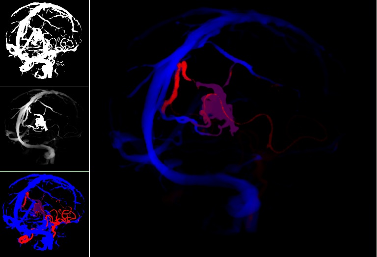
Cerebral arteriovenous malformations (AVMs) are a type of vascular anomaly consisting of large intertwined vascular growth (the nidus) that are prone to serious hemorrhaging and can result in patient death if left untreated. Intervention through surgical clipping of feeding and draining vessels to the nidus is a common treatment. However, identification of which vessels to clip is challenging even to experienced surgeons aided by conventional image guidance systems. In this work, we describe our methods for processing static preoperative angiographic images in order to effectively visualize the feeding and draining vessels of an AVM nidus. Maps from level-set front propagation processing of the vessel images are used to label the vessels by colour. Furthermore, images are decluttered using the topological distances between vessels. In order to aid the surgeon in the vessel clipping decision-making process during surgery, the results are displayed to the surgeon using augmented virtuality.
Decluttering and visualizing blood flow
Publications
- S. J. S. Chen, M. Kersten-Oertel, S. Drouin, and D. L. Collins. “Visualizing the path of blood flow for image guided surgery of cerebral arteriovenous malformations”. SPIE Medical Imaging, San Diego, CA, Feb 4–9, 2012.
- M. Kersten-Oertel, S. J. S. Chen, D. L. Collins. “Enhancing depth perception of volume-rendered angiography data”.VIS 2011 Poster Session, Providence, RI, Oct. 23–38, 2011.