Multi-contrast PD25 atlas


images-NIST.003
This set of multi-contrast population-averaged PD brain atlas contains 5 different image contrasts:  T1w ( FLASH & MPRAGE), T2*w, T1–T2* fusion, phase, and an R2* map. Probabilistic tissue maps of whiter matter, grey matter, and cerebrospinal fluid are provided for the atlas. We also manually segmented eight subcortical structures: caudate nucleus, putamen, globus pallidus internus and externus (GPi & GPe), thalamus, STN, substantia nigra (SN), and the red nucleus (RN). Lastly, a co-registered histology-derived digitized atlas containing 123 anatomical structures is included.
 
segmentation-demo1-s2.0-S2352340917301452-gr5
We employed a novel T1–T2* fusion MRI that visualizes both cortical and subcortical structures to drive groupwise registration to create co-registered multi-contrast  unbiased templates from 25 PD patients that later went for the STN deep brain stimulation procedure. The finished atlas is in ICBM152 space. Three different resolutions are provided: 1×1×1 mm, 0.5×0.5×0.5 mm, and sectional 0.3×0.3×0.3 mm.

The included files are as followed:
R2* map: PD25-R2starmap-atlas-{0.3mm, 0.5mm, 1mm}
phase map: PD25-phase-atlas-{0.3mm, 0.5mm, 1mm}
MPRAGE T1: PD25-T1MPRAGE-template-{0.3mm, 0.5mm. 1mm}
FLASH T1: PD25-T1GRE-template-{0.3mm, 0.5mm, 1mm}
T2*w: PD25-T2star-template-{0.3mm, 0.5mm, 1mm}

T1-T2* fusion: PD25-fusion-template-{0.3mm, 0.5mm, 1mm}

Brain masks: PD25-atlas-mask-{0.3mm, 0.5mm, 1mm}
Probabilistic brain tissue maps: PD25-{WM,GM,CSF}-tissuemap
8 subcortical structure segmentation: PD25-subcortical-1mm
High resolution midbrain nuclei manual segmentation: PD25-midbrain-0.3mm

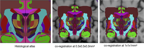
Co-registered histological atlas:  PD25-histo-{0.3mm, 1mm}

midbrain labels: PD25-midbrain-labels.csv
Subcortical labels: PD25-subcortical-labels.csv
Histological labels: PD25-histo-labels.csv


BigBrain co-registration

To help bridge the insights of micro and macro-levels of the brain, the Big Brain atlas was nonlinearly registered to the PD25 and ICBM152 (symmetric and asymmetric) atlases in a multi-contrast registration strategy, and subcortical structures were manually segmented for BigBrain, PD25 , and ICBM152 atlases. To help relate PD25 atlas to clinical T2w MRI, a synthetic T2w PD25 atlas was also created. The registered BigBrain atlases are available at the resolutions of 1×1×1 mm, 0.5×0.5×0.5 mm, and 0.3×0.3×0.3 mm.
Screen Shot 2019-02-23 at 11.06.48 PM
Data related to BigBrain co-registration:

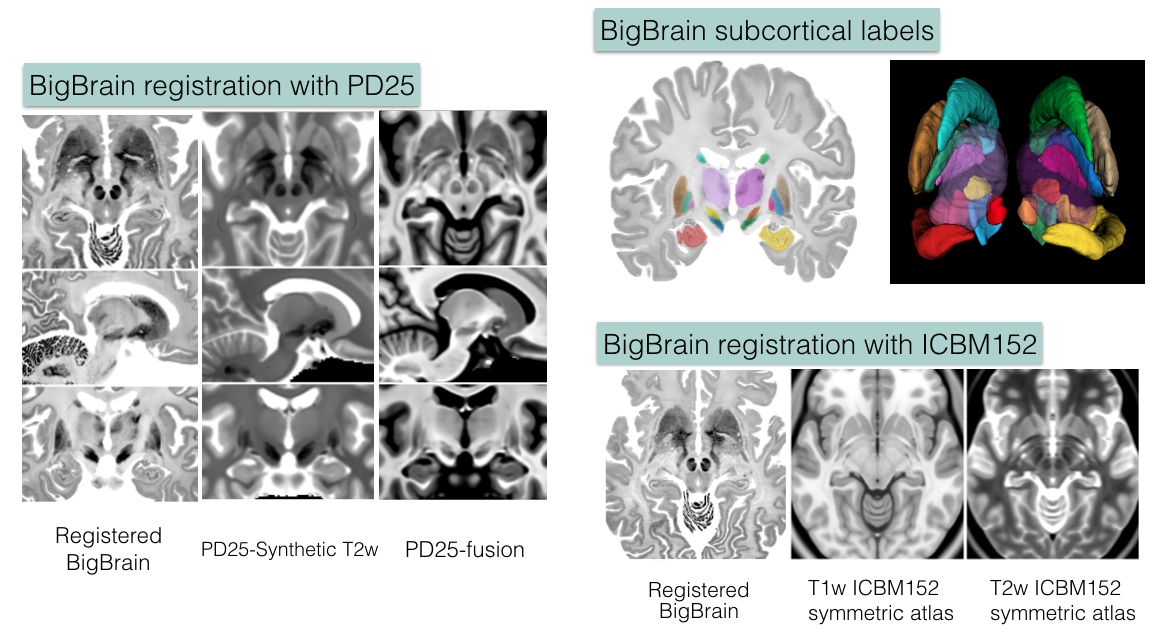
1. Deformed BigBrain atlases:

  • BigBrain in PD25 space: BigBrain-to-PD25-nonlin-{300um, 0.5mm, 1mm}
  • BigBrain in ICBM152 symmetric atlas: BigBrain-to-ICBM2009sym-nonlin-{300um, 0.5mm, 1mm}
  • BigBrain in ICBM152 asymmetric atlas: BigBrain-to-ICBM2009asym-nonlin-{300um, 0.5mm, 1mm}
  • Synthetic T2w PD25 atlas: PD25-SynT2-template-{300um, 0.5mm, 1mm}
  • T1-T2* fusion PD25 atlas: PD25-enhanceFusion-template-{300um, 0.5mm, 1mm}

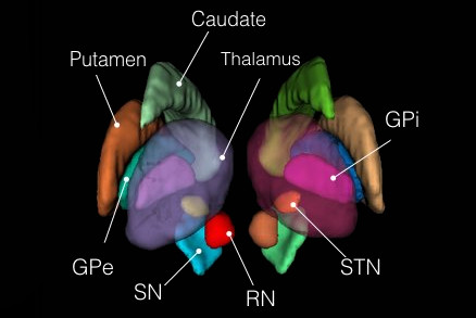
2. Manual subcortical segmentations:

  • BigBrain coregistered to ICBM in the BigBrain2015 release: BigBrain-segmentation-0.3mm
  • MNI PD25: PD25-segmentation-0.5mm
  • ICBM152 2009b symmetric: ICBM2009b_sym-segmentation-0.5mm
  • ICMB152 2009b asymmetric: ICBM2009b_asym-segmentation-0.5mm

3. Related transformations:

  • BiBrain-to-PD25: BigBrain-to-PD25-nonlin.xfm
  • BigBrain-to-ICBM2009asym: BigBrain-to-ICBM2009asym-nonlin.xfm
  • BigBrain-to-ICBM2009sym: BigBrain-to-ICBM2009sym-nonlin.xfm
  • PD25-to-ICBM2009asym: PD25-to-ICBM2009asym-nonlin.xfm
  • PD25-to-ICBM2009sym: PD25-to-ICBM2009sym-nonlin.xfm

4. List of subcortical labels: subcortical-labels.csv


Publications

For the methods used, and to use the atlas for research purposes, please cite the following articles:
  1. Y. Xiao, V. Fonov, S. Beriault, F.A. Subaie, M.M. Chakravarty, A.F. Sadikot, G. Bruce Pike, and D. Louis Collins, “A dataset of multi-contrast population-averaged brain MRI atlases of a Parkinson’s disease cohort,” accepted in Data in Brief, 2017.
  2. Y. Xiao, V. Fonov, S. Beriault, F.A. Subaie, M.M. Chakravarty, A.F. Sadikot, G. Bruce Pike, and D. Louis Collins, “Multi-contrast unbiased MRI atlas of a Parkinson’s disease population,” International Journal of Computer-Assisted Radiology and Surgery, vol. 10(3), pp. 329-341, 2015.
  3. Y. Xiao, S. Beriault, G. Bruce Pike, and D. Louis Collins, “Multicontrast multiecho FLASH MRI for targeting the subthalamic nucleus,” Magnetic Resonance Imaging, vol. 30, pp. 627-640, 2012.

If you are using the BigBrain atlas co-registration dataset, please refer to the following preprint:

  1. Y. Xiao, J.C. Lau, T. Anderson, J. DeKraker, D. Louis Collins, T. Peters, and A.R. Khan, “Bridging micro and macro: accurate registration of the BigBrain dataset with the MNI PD25 and ICBM152 atlases,” 

If you are using the Big Brain data, please cite the following publication:

  1. Amunts, K. et al.: “BigBrain: An Ultrahigh-Resolution 3D Human Brain Model”, Science (2013) 340 no. 6139 1472-1475, June 2013

Copyright

Copyright (C) 2016,2017,2018 Yiming Xiao, McConnell Brain Imaging Centre,
Montreal Neurological Institute, McGill University.

License

PD25 atlases are distributed under CC BY-NC-SA 3.0 Licence

Dataset for BigBrain co-registration with PD25 and ICBM152 is under CC BY 4.0 Licence. Note that this exception to the existing BigBrain dataset does not alter the general term of the license for the use of BigBrain itself, which is still under CC BY-NC-SA 4.0 License.

Download

Version 20170213: Download archives containing brain atlases, brain masks, midbrain and subcortical segmentation and histological labels: MINC1, MINC2, NIFTI

Version 20160706: Download archives containing brain atlases, brain masks and midbrain segmentation: MINC1, MINC2, NIFTI

Co-registration of BigBrain with PD25 and ICBM152 atlases: Download archives containing registered Big Brain atlas, manual segmentations, and registration transformation (only available in MINC2 package): MINC2, NIFTI

Ultrasound and Augmented Reality

For intraoperative use, neuronavigation systems must relate the physical location of a patient with the preoperative models by means of a transformation that relates the two through a patient-to-image mapping. By tracking the patient and a set of specialized surgical tools, this mapping allows a surgeon to point to a specific location on the patient and see the corresponding anatomy on the patient specific models. However, throughout the intervention, hardware movement, an imperfect patient-image mapping, and movement of brain tissue during surgery invalidates the patient-to-image mapping. These sources of inaccuracy, collectivey described as ‘brain shift’, reduce the effectiveness of using preoperative patient specific models intraoperatively. Additionally, the surgeon is left with the cognitive burden of merging the virtual models of the patient with the visible and invisible physical anatomy.

An underlying advantage of IBIS (IBIS Neuronav) is that it allows for both individual streams of research as well as the combination of different streams to overcome major or minor pitfalls within them. This is demonstrated through our combination of iUS and AR for improving the accuracy of AR visualizations during tumour neurosurgeries. With this combination of technologies, the interpretation difficulties associated with US images are mediated with detailed AR visualizations and the accuracy issues associated with AR are corrected through registration of the US images. This allows for improved patient-specific planning intra-operatively by both prolonging the reliable use of neuronavigation and the understanding of complex three dimensional medical imaging data so that different surgical strategies can be adapted when necessary.

The avatar represents the orientation of the patient’s head. The surgical field of view (left), the AR view before US correction where the tumour seems to conform unnaturally to the surrounding tissue (middle), and the brain shift corrected AR view where the tumour visualization now lines up naturally with surrounding tissue and can be used for accurate intra-operative planning.

Publications

[1] Gerard, Ian J., Marta Kersten-Oertel, Simon Drouin, Jeffery A. Hall, Kevin Petrecca, Dante De Nigris, Tal Arbel, and D. Louis Collins. “Improving Patient Specific Neurosurgical Models with Intraoperative Ultrasound and Augmented Reality Visualizations in a Neuronavigation Environment.” InClinical Image-Based Procedures. Translational Research in Medical Imaging, pp. 28-35. Springer International Publishing, 2015.

[2] Gerard, Ian J., Marta Kersten-Oertel, Simon Drouin, Jeffery A. Hall, Kevin Petrecca, Dante De Nigris, Tal Arbel, and D. Louis Collins. “Improving Augmented Reality Tumour Visualization With Intraoperative Ultrasound In Image Guided Neurosurgery: Case Report.” International Journal of Radiology and Surgery 10(S1):1-312, 2015.


Medial Temporal Lobe (MTL) Segmentation

Our group has developed multiple accurate segmentation methods during years. Our nonlinear patch-based method with error correction is one of the most accurate methods of segmentation of medial temporal lobe structures. Our method reaches 0.901 of Dice similarity with manual tracing for hippocampus which is one of the highest values reported in the literature. Medial temporal lobe atrophy and especially hippocampal atrophy has been one of the most investigated AD biomarkers.

  1. Zandifar, A., Fonov, V. , Coupé, P., Pruessner, J. C.,  & Collins, D. L. (2014). A unified assessment of fully automated hippocampus segmentation methods. Alzheimer’s & Dementia: The Journal of the Alzheimer’s Association10(4), P86.
  2. Zandifar, A., Fonov, V. , Coupé, P., Pruessner, J. C.,  & Collins, D. L. (2015). A quantitative comparison between two manual hippocampal segmentation protocolsAlzheimer’s & Dementia: The Journal of the Alzheimer’s Association, 11(7), P67-P68.

Spinal Surgery

Each year more than 20,000 Canadians are treated surgically for lower back pain. Image guided surgical (IGS) techniques can reduce the number of complications that can arise in classical surgical techniques that can be as high as 20–30%. Intraoperative imaging can further reduce risks by improving accuracy and precision in the placement of pedicular screws used for lumbar fixation. This project will investigate the use of intra-operative ultrasound as an inexpensive alternative to intraoperative CT or MRI. We will develop techniques to precisely identify the boney surface of the vertebrae in both CT and ultrasound and use this information to improve patient-image registration required for image guided spine surgery. This in turn will improve the accuracy in guidance and precision of screw placement and result in better care for the patient.

We have tested the hypothesis that intra-operative ultrasound is viable, precise and clinically relevant to improved precision and will reduce operating time in image guided surgery of the spine. Our specific aims were:

  • To develop an automated model-based method to segment vertebrae of the human spine from 3D computed tomography data and ultrasound data.
  • To develop an automated slice-to-volume registration method using intraoperative 2D ultrasound (US) to align a patient’s vertebra to pre-operative CT images to improve the accuracy of image guided surgery and to reduce the time required for registration.
  • To validate and characterized the accuracy of the registration and segmentation methods in vitro using human plastic spine models, swine models and human cadaver specimens.
  • To evaluate the precision and speed of the ultrasound-based registration method in vivo with patients in the context of lumbo-sacral pedicle screw implantation with respect to landmark-based registration in a commercial navigation system.

Increasing the accuracy of the patient-image registration enabled the surgeon to improve pedicular screw positioning and thus decrease risks for the spinal cord, nerve roots or blood vessels. Improved precision can also increase instrumentation strength, thus preventing loosening of the misplaced hardware. When used with an anterior approach, the IGS system can be used to facilitate removal of disks or tumors, or positioning of artificial disks. By decreasing the time required for the registration procedure, overall operating time will be reduced, implying shorter muscle retraction times with the potential of reduced post-operative pain and reduced risk of infection for the patient. For the clinical team, shorter operating times will reduce fatigue, and may enable completion of more complex procedures with greater assurance. This work was supported by a CIHR operating grant (PI: Collins, co-PIs: Goulet).

References

[1] Fonov VS, Le Troter A, Taso M, De Leener B, Lévêque G, Benhamou M, Sdika M, Benali H, Pradat PF, Collins DL, Callot V, Cohen-Adad J. Framework for integrated MRI average of the spinal cord white and gray matter: The MNI-Poly-AMU template. Neuroimage. 2014 Sep 7;102P2:817–827

[2] G Forestier, F Lalys, DL Collins, J. Meixensberger, S Wassef, T Neumuth, B Goulet, L Riffaud, P Jannin. Multi-site study of surgical practice in neurosurgery based on Surgical Process Models, Journal of Biomedical Informatics, 46(5), October 2013, Pages 822–829

[3] Yan CX, Goulet B, Chen SJ, Tampieri D, Collins DL. Validation of automated ultrasound-CT registration of vertebrae. Int J Comput Assist Radiol Surg. 2012 Jul;7(4):601–10

[4] Yan, C. X., Goulet, B., Tampieri, D., & Collins, D. L. (2012). Ultrasound-CT registration of vertebrae without reconstruction. International journal of computer assisted radiology and surgery, 7(6), 901–909.

[5] C.X.B. Yan, B. Goulet, J. Pelletier, S.J.S. Chen, D. Tampieri and D.L. Collins, “Towards Accurate, Robust and Practical Ultrasound-CT Registration of Vertebrae for Image-Guided Spine Surgery,” International Journal of Computer Assisted Radiology and Surgery, 2011 Jul;6(4):523–37.

Schizophrenia

The NIST Lab has collaborated with the Utrecht group headed by Dr. H. Hulshoff Pol for the past number of years [1, 2, 3]. Her group has used my ANIMAL non-linear registration software with voxel based morphometry (VBM) for morphometric analysis of cerebral MRI of patients with schizophrenia. Recently they have provided evidence which suggests that brain structures reflect overlapping and segregating genetic liabilities for schizophrenia and BD. The overlapping smaller white matter volume and common areas of thinner and/or thicker cortex (thinner right orbitofrontal cortex, thinner right (and left) parahippocampus, and thicker temporoparietal and left superior motor cortices) suggest that both disorders share neurodevelopmental roots. They have also demonstrated excessive thinning of the cortex over time, most pronounced in the temporal and frontal areas, with progression across the span of the illness. This excessive thinning appears related to medication and outcome. Their findings were extended to reveal that cannabis use in patients with schizophrenia resulted in additional thinning in the left dorsolateral prefrontal cortex (DLPFC), left anterior cingulate cortex (ACC) and left occipital lobe as compared to those patients with schizophrenia that did not use cannabis after a 5-year follow-up.

In later papers, they have identified focal grey-matter differences in a number of regions when compared to age-matched controls [4]. They have also found focal white matter changes that suggest abnormal inter-hemispheric connectivity of anterior cortical and sub-cortical brain regions, reflecting decreased hemispheric specialisation in schizophrenia [5]. These results have been partially replicated in a twin study [6,7] and a long term follow-up study [8].

References

[1] Hulshoff Pol HE, van Baal GC, Schnack HG, Brans RG, van der Schot AC, Brouwer RM, van Haren NE, Lepage C, Collins DL, Evans AC, Boomsma DI, Nolen W, Kahn RS. Overlapping and segregating structural brain abnormalities in twins with schizophrenia or bipolar disorder. Arch Gen Psychiatry. 2012 Apr;69(4):349–59

[2] van Haren, Neeltje EM, Hugo G. Schnack, Wiepke Cahn, Martijn P. van den Heuvel, Claude Lepage, Louis Collins, Alan C. Evans, Hilleke E. Hulshoff Pol, and René S. Kahn. “Changes in cortical thickness during the course of illness in schizophrenia.” Archives of general psychiatry. 2011; 68(9): 871–880.

[3] M. Rais, N.E.M. van Haren, W Cahn, H.G. Schnack, C. Lepage, D.L. Collins, A. Evans, H. E. Hulshoff Poll and R.S. Kahn, “Cannabis use and progressive cortical thickness loss in areas rich in CB1 receptors during the first five years of schizophrenia,” European Neuropsychopharmacology, 2010 Dec; 20(12): 855–65.

[4] H. E. Hulshoff Pol, H. G. Schnack, R. C. Mandl, N. E. van Haren, H. Koning, D. L. Collins, A. C. Evans, and R. S. Kahn, “Focal gray matter density changes in schizophrenia,” Arch Gen Psychiatry. 2001; 58: 1118–25.

[5] H. E. Hulshoff Pol, H. G. Schnack, R. C. Mandl, W. Cahn, D. L. Collins, A. C. Evans, and R. S. Kahn, “Focal white matter density changes in schizophrenia: reduced inter-hemispheric connectivity,” Neuroimage. 2004; 21: 27–35.

[6] H. Hulshoff Pol, H. G. Schnack, R. C. Mandl, R. G. H. Brans, N. E. van Haren, W. F. C. Baare, C. J. van Oel, D. L. Collins, A. C. Evans, and R. S. Kahn, “Gray and white matter density changes in monozygotic and same-sex dizygotic twins discordant for schizophrenia: a voxel-based morphometry study,” presented at 10th Annual Meeting of the Organization for Human Brain Mapping, Budapest, Hungary, 2004.

[7] H. E. Hulshoff Pol, H. G. Schnack, R. C. Mandl, R. G. Brans, N. E. van Haren, W. F. Baare, C. J. van Oel, D. L. Collins, A. C. Evans, and R. S. Kahn, Gray and white matter density changes in monozygotic and same-sex dizygotic twins discordant for schizophrenia using voxel-based morphometry, Neuroimage. 2006; 31(2): 482–8.

[8] N. E. van Haren, H. E. Hulshoff Pol, H. G. Schnack, W. Cahn, R. C. Mandl, D. L. Collins, A. C. Evans, and R. S. Kahn, “Focal Gray Matter Changes in Schizophrenia across the Course of the Illness: A 5-Year Follow-Up Study,” Neuropsychopharmacology. 2007; 32: 2057–66.

Brain Shift

Since the introduction of the first intraoperative frameless stereotactic navigation device, image guided neurosurgery has become an essential tool for many neurosurgical procedures due to its ability to minimize surgical trauma by allowing for the precise localization of surgical targets. The integration of preoperative image information into a comprehensive patient-specific model enables surgeons to preoperatively evaluate the risks involved and define the most appropriate surgical strategy. Perhaps more importantly, such systems enable surgery of previously inoperable cases by helping to locate safe surgical corridors through IGNS-identified non-critical areas.

For intraoperative use, neuronavigation systems must relate the physical location of a patient with the preoperative models by means of a transformation that relates the two through a patient-to-image mapping. Throughout the intervention, hardware movement, an imperfect patient-image mapping, and movement of brain tissue during surgery invalidates the patient-to-image mapping. These sources of inaccuracy, collectivey described as ‘brain shift’, reduce the effectiveness of using preoperative patient specific models intraoperatively. Intraoperative imaging, such as MRI, has been shown to improve the accuracy of tumour resections through lengthened image guidance. However, such technology is extremely expensive, prolongs surgery, poses logistical challenges during awake surgeries, and is available in only a few centres worldwide. We have developed a neuronavigation platform (IBIS Neuronav) that integrates tissue deformation tracking during surgery based on tracked intraoperative ultrasound (iUS) that can accurately align all pre-operative data to the iUS to account for brain shift throughout a surgical intervention.


Reference:

[1] I. Gerard and D. L. Collins, “An Analysis of Tracking Error in Image Guided Neurosurgery”, Int. J. Computer Assisted Radiolgy and Surgery. 2015, Jan 4; 1–10 [Epub ahead of print].

[2] H. Rivaz, D.L. Collins, “Near real-time robust non-rigid registration of volumetric ultrasound images for neurosurgery”, Ultrasound in Medicine and Biology. 2015 Feb; 41(2): 574–587.

[3] H. Rivaz, S.J.S Chen, D.L. Collins, “Automatic Deformable MR-Ultrasound Registration for Image-Guided Neurosurgery”, IEEE Transactions on Medical Imaging. 2015 Feb; 34(2); 366–380.

[4] H. Rivas, Z. Karimaghaloo, D.L. Collins, “Nonrigid Registration of Ultrasound and MRI Using Contextual Conditioned Mutual Information”, IEEE Trans Med Imag. 2014 Mar;33(3):708–25.

[5] S. Beriault, A. Sadikot, F. Alsubaie, S. Drouin, D.L. Collins, G.B. Pike. “Neuronavigation using susceptibility-weighted venography: application to deep brain stimulation and comparison with gadolinium contrast”, Journal of Neurosurgery. 2014 Jul;121(1):131–41.

[6] L. Mercier, D Araujo, C Haegelen, RF Del Maestro, K Petrecca, DL Collins, “Registering pre- and post-resection 3D ultrasound for improved residual brain tumor localization”, Ultrasound in Medicine and Biology, 2013 Jan;39(1):16–29.

[7] M. Kersten-Oertel, P. Jannin, D.L. Collins, “The State of the Art in Mixed Reality Visualization in Image-Guided Surgery”, IEEE Transactions on Visualization and Computer Graphics. 2013 Mar;37(2):98–112.

[8] D. De Nigris, D. L. Collins, T. Arbel, “ Fast Rigid Registration of Pre-Operative Magnetic Resonance Images to Intra-Operative Ultrasound for Neurosurgery based on High Confidence Gradient Orientations”, 2013 July; 8(4): 649–661.


Epilepsy

In some patients with refractory epilepsy that are candidates for surgery, intracranial EEG is recorded to precisely localize the epileptic focus. To record intracranial EEG, multiple depth electrodes are surgically implanted through holes in the skull, each with 8–15 equally spaced contacts. Current implantation planning consists of visual inspection of the patient’s MRI & CTA, visually searching for paths to the targets while avoiding vessels. This procedure is sub-optimal since estimation of the number of electrodes needed to sample a region and the precise location of each contact, which is paramount to accurately identify the focus and its extent, cannot be considered.

The goal of this project, conducted by Dr. Zelmann, one of my postdoctoral fellows, is to evaluate the clinical use of optimized depth electrode planning. We hypothesize that the use of our computed aided procedure will increase the accuracy and amount of information obtained during EEG intracranial investigation while constraining the trajectories to safe paths.

We have developed a procedure to optimize electrode location that would enable us to obtain complete coverage of a lesion or region of interest and surrounding neocortical grey matter, while minimizing the risk of approaching vessels and other critical structures. To this end, we automatically segment MRI, CT and CTA data; we model each electrode as a cylinder to assess risk; we estimate the contribution of individual contacts to record EEG; we compute an aggregated score for each electrode and a global score, obtaining the best cohort as the combined set of electrodes that remain at a safe distance.

To allow its clinical use during planning and surgical implantation, the procedure is integrated into an image-guided neuronavigation system developed in our research laboratory over the past decade, which is called IBIS (Interactive Brain Imaging System; Mercier et al., Int J CARS. 2011;6(4):507–522). IBIS allows planning and navigation based on preoperative medical image data. The best cohort and a list of possible electrodes per target (ordered in terms of risk and recorded EEG) is generated and displayed in IBIS and will be available during planning and in the OR. The surgeon can visually review trajectories, decide between one of the automatic trajectories and the manual one and, if necessary, re-plan a trajectory in almost real time.

Preliminary results [1] presented at an international workshop on clinical image-based procedures, suggest that automatic planning allows recording from a larger volume than manually planned trajectories (p<0.01) and from more temporal grey matter (p<0.001), while remaining further away from segmented vessels (p<0.01). We are currently evaluating the procedure in retrospective clinically acquired imaging data from 20 patients with electrodes implanted in MTL regions. We are comparing visual and automatic trajectories by estimating volume recording from the target volumes (in this case: amygdala and hippocampus) and neocortical temporal grey matter as well as distance to vessels and other critical structures.

Reference

[1] Zelmann, R., Beriault, S., Mok, K., Haegelen, C., Hall, J., Pike, G. B., Olivier, A & Collins, D. L. (2014). Automatic Optimization of Depth Electrode Trajectory Planning. In Clinical Image-Based Procedures. Translational Research in Medical Imaging (pp. 99–107). Springer International Publishing.

[2] R. Zelmann, S. Beriault, K. Mok, C. Haegelen, J. Hall, G.B. Pike, A. Olivier & D.L. Collins, “Improving Recorded Volume in Mesial Temporal Lobe by Optimizing Stereotactic Intracranial Electrode Implantation Planning” submitted to International Journal of Computer Assisted Radiology and Surgery (CARS-D-14–00251), Oct 9, 2014.

[3] Zelmann, R., Beriault, S., Mok, K., Haegelen, C., Hall, J., Pike, G. B., Olivier, A & Collins, D. L. (2014). Automatic Optimization of Depth Electrode Trajectory Planning. In Clinical Image-Based Procedures. Translational Research in Medical Imaging (pp. 99–107). Springer International Publishing.

[4] R. Zelman, D.L. Collins, “Automatic Optimization of Depth Electrode Trajectory Planning”, MICCAI 2013 Workshop on Clinical Image-based Procedures: Translational Research in Medical Imaging

[5] C. Haegelen, P. Perucca, C.-E. Châtillon, L. Andrade-Valença, R. Zelmann, J. Jacobs, D. L. Collins, F. Dubeau, A. Olivier and J. Gotman. “High-frequency oscillations, extent of surgical resection and surgical outcome in drug-resistant focal epilepsy”, Epilepsia 2013 May;54(5):848–57.

[6] Zelmann, R., S. Beriault, M. M. Marinho, K. Mok, J. A. Hall, N. Guizard, C. Haegelen, A. Olivier, G. B. Pike, and D. L. Collins. “Improving recorded volume in mesial temporal lobe by optimizing stereotactic intracranial electrode implantation planning.” International journal of computer assisted radiology and surgery (2015): 1–17.

Multiple Sclerosis

Multiple sclerosis (MS) is a neurological disease that predominately affects young adults. Inflammatory mechanisms were believed to be the main contributor to the development of MS. However, more recent neuropathological and magnetic resonance imaging (MRI) studies suggest that neurodegenerative processes play an equivalent central role, that these degenerative processes commence during the early stages of the disease, and that neuronal and axonal loss may be the key substrate for the development of disability. One macroscopic hallmark of neurodegeneration is brain atrophy, which can be readily investigated non-invasively using MRI. We have recently demonstrated brain atrophy in pediatric-onset MS patients, further supporting a very early and possibly primary role for neurodegeneration in MS.

The three current hypotheses for the specific pathobiology underlying brain atrophy in MS are:

  • 1) atrophy in focal lesions and normal-appearing brain tissue occurs secondary to neuroaxonal injury and Wallerian degeneration and loss of normal myelin or reduction in myelin density associated with inflammation-mediated tissue insult;
  • 2) atrophy is due to a diffuse, primary degenerative process associated with neuronal cell death and subsequent loss of axons and myelinated pathways; and
  • 3) atrophy is due to a combination of mechanisms 1 and 2. In children with MS, insult to precursors of primary myelination may further impede normal brain maturation contributing to failure of age-expected brain growth in addition to atrophic loss of established neural networks.

The onset of MS during childhood and adolescence provides a potentially enhanced capacity to distinguish the earliest aspects of MS pathobiology, as the young age of such patients inherently limits the time period available for subclinical disease. A further unique aspect of pediatric onset MS is the potential deleterious impact of MS pathobiology to the processes of primary myelination and normal brain maturation, and thus potential consequence of MS contributing to failure of age-expected brain growth. We have recently reported reduced brain volumes in pediatric-onset MS compared to pediatric normal controls in cross-sectional studies. We also noted that the reduction in brain volume was not only global, but also specifically notable in the thalamus. We now propose to delineate whether the reduced brain volumes reflect age-expected failure of normative growth or loss of previously developed brain tissue (atrophy), or both, and will further explore the selective vulnerability of specific brain regions.

MRI confirmation of progressive brain volume loss, detectable in children and adolescents with MS, will not only refute the concept of pediatric brain resiliency and enhanced repair, but will also emphasize the fundamental nature of neurodegenerative biology of MS. Such confirmation has significant import on future therapeutic strategies, as it implies that anti-inflammatory therapies alone may fail to mitigate the negative impact of MS, and that neuroprotective strategies will be required from onset. This study is supported by an operating grant (Biomedical Research) provided by the Multiple Sclerosis Society of Canada & The Multiple Sclerosis Scientific Research Foundation.

Reference

[1] R. Harmouche, N.K. Subbanna, D.L. Collins, D.L. Arnold, T. Arbel, “Probabilistic multiple sclerosis lesion classification based on modeling regional intensity variability and local neighbourhood information”, IEEE Transactions on Biomedical Engineering, 2015 May; 62(5): 1281–92.

[2] N Guizard, P Coupé, VS Fonov, JV Manjón, DL Arnold, DL Collins, “Rotation-invariant multi-contrast non-local means for MS lesion segmentation (RMNMS)”, NeuroImage: Clinical. 2015 May 8: 376–89.

[3] K. Weier, B. Banwell, A. Cerasa, D.L. Collins, A. Dogonowski, H. Lassmann, A. Quattrone, M.A. Sahraian, H.R. Siebner and T. Sprenger, “The role of the cerebellum in multiple sclerosis”, Cerebellum. 2015 Jun; 14(3); 364–74

[4] Aubert-Broche B, Fonov VS, Garcia-Lorenzo D, Mouiha A, Guizard N, Coupé P, Eskildsen SF, Collins DL. A new method for structural volume analysis of longitudinal brain MRI data and its application in studying the growth trajectories of anatomical brain structures in childhood. Neuroimage 2013;82C:393–402.

[5] Aubert-Broche B, Fonov V, Ghassemi R, Arnold DL, Banwell B, Sled JG, Collins DL. Regional brain atrophy in children with multiple sclerosis. Neuroimage 2011;58:409–415.

[6] Kerbrat A, Aubert-Broche B, Fonov V, Narayanan S, Sled JG, Arnold DL, Banwell B, Collins DL. Reduced head and brain size for age and disproportionately smaller thalami in child-onset MS. Neurology 2012;78:194–201.

Parkinson’s Disease

Parkinson’s disease (PD) is the second most common neurodegenerative disease, after Alzheimer’s disease, worldwide. At the NIST Lab, we are dedicated to explore potential structural biomarkers for the disease, as well as proposing image processing techniques for the surgical treatment of PD.


Projects include:

Stereotaxic surgery for movement disorders

Stereotaxic surgery for movement disorders

We have recently developed techniques [1] used to create a lower resolution 3D atlas, based on the Schaltenbrand and Wahren print atlas, which was integrated into a stereotactic neurosurgery planning and visualization platform (VIPER), and a higher resolution 3D atlas derived from a single set of manually segmented histological slices containing nuclei of the basal ganglia, thalamus, basal forebrain, and medial temporal lobe. We have therefore developed, and are continuing to validate, a high-resolution computerized MRI-integrated 3D histological atlas, which is useful in functional neurosurgery, and for functional and anatomical studies of the human basal ganglia, thalamus, and basal forebrain.

Parkinson’s disease (PD) is a neurodegenerative disorder that impairs the motor functions. Deep brain stimulation (DBS) is an effective therapy to treat drug-resistant PD. Accurate placement of the DBS electrode deep in the brain under stereotaxic conditions is key to successful surgery [2]. Accuracy depends on a number of factors including registration error of the stereotaxic frame, geometric distortion of the MRI scans and brain tissue shift during resulting from cerebrospinal fluid (CSF) leakage, cranial pressure change, and gravity after the burr-hole is opened.

By scanning through acoustic skull windows, transcranial ultrasound can provide non-invasive visualization of internal brain structures (i.e. midbrain, blood vessels, and certain nuclei like the substantia nigra) as well as metallic surgical instruments (i.e. DBS electrode and cannula). We believe that such images can be used to improve stereotaxic accuracy.

In the past decade, we have developed a prototype image-guided neuronavigation system called IBIS (Interactive Brain Imaging System) in our research laboratory, which enables the acquisition of intraoperative 2D/3D ultrasound, and addresses the issue of registration errors caused by brain shift by using ultrasound data to improve the patient/image alignment. By linking the preoperative MRI, and the corresponding surgical plan, to the transcranial ultrasound with appropriate registration methods, we will enable real-time monitoring of the DBS implantation and will improve the safety and accuracy of the procedure. Our goal is to acquire transcranial ultrasound images, and examine its performance as an intraoperative imaging modality.

The study, as well as surgical treatment of PD necessitate the delineation of basal ganglia nuclei morphology. Few automatic volumetric segmentation methods have attempted to identify the key brainstem substructures including the subthalamic nucleus (STN), substantia nigra (SN), and red nucleus (RN) due to their small size and poor contrast in conventional MRI. I recently developed a technique [3] with my Ph.D. student Yimming Xiao based on a dual-contrast patch-based label fusion method to segment the SN, STN, and RN. Our proposed method outperformed the state-of-the-art single-contrast patch-based method for segmenting brainstem nuclei using a multi-contrast multi-echo FLASH MRI sequence. This method is encouraging as it will provide promising developments for the treatment and research of PD. This study is supported by the NSERC/CIHR Collaborative Health Research Program

Reference

[1] A. F. Sadikot, M. M. Chakravarty, G. Bertrand, V. V Rymar, F. Al-Subaie and D. L. Collins. “Creation of computerized 3D MRI-integrated atlases of the human basal ganglia and thalamus”, Frontiers in Systems Neuroscience, 2011;5:71

[2] Y. Xiao, V.S. Fonov, S. Beriault, F. Al Soubaie, M.M. Chakravarty, A.F. Sadikot, G.B. Pike and D.L. Collins, “Multi-contrast unbiased MRI atlas of a Parkinson’s disease population”, Int J Comput Assist Radiol Surg. 2015 March; 10(3):329–41.

[3] Xiao Y, Fonov VS, Beriault S, Gerard I, Sadikot AF, Pike GB, Collins DL. Patch-based label fusion segmentation of brainstem structures with dual-contrast MRI for Parkinson’s disease. Int J Comput Assist Radiol Surg. 2015 July; 10(7):1029–41

[4] M.M. Chakravarty, G. Bertrand, C. Hodge, A.F. Sadikot, and D.L. Collins, “The creation of a brain atlas for image guided neurosurgery using serial histological data,” NeuroImage. 2006; 30(2): 359–76.

YouTube-logo-full_color