VentRa

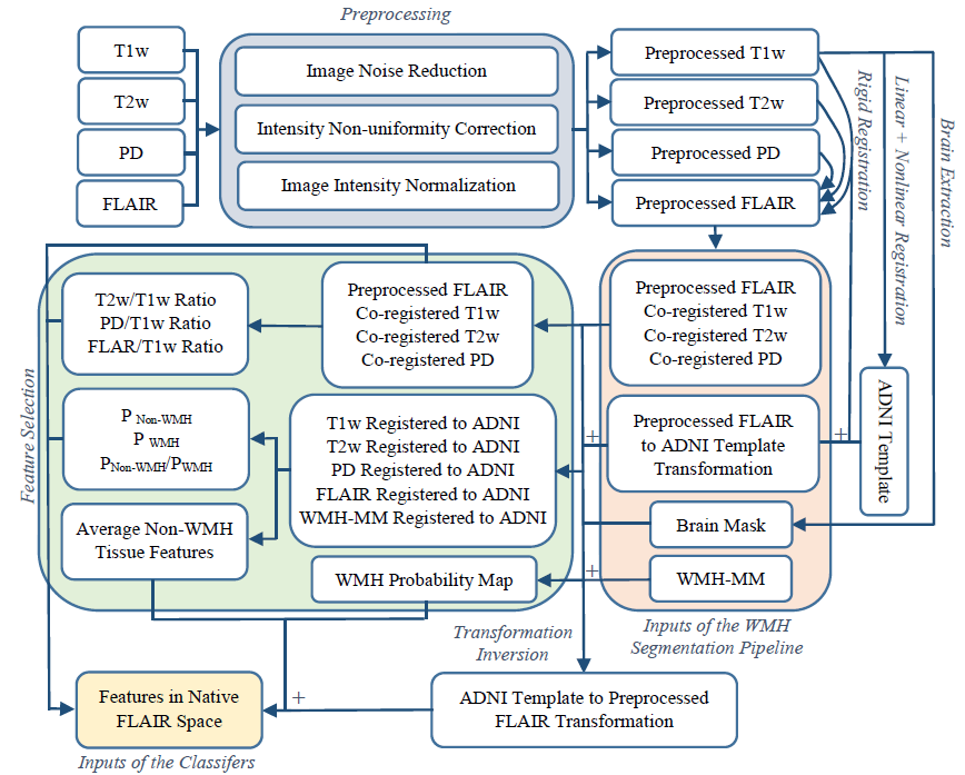
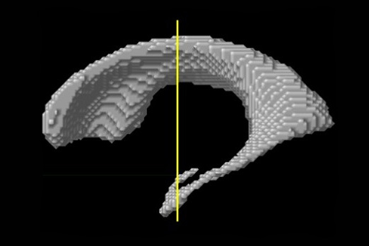
Lateral ventricles are reliable and sensitive indicators of brain atrophy and disease progression in behavioral variant frontotemporal dementia (bvFTD). VentRa takes a comma separated (.csv) file providing the path for the raw T1-weighted images as well as age and sex of the subjects as input, and provides preprocessed images along with ventricle segmentations, QC files for the segmentations, as well as a .csv file including the diagnosis (based on the classifier trained on bvFTD vs the mixed group data) along with all the extracted ventricle features: i.e. total ventricle volume, ventricle volumes in each lobe and hemisphere,anterior-posterior ratio (APR), left-right temporal lobe ratio (LRTR), and left-right frontal ratio (LRFR).

ventra

VentRa Tool

For more details, see:

Ana L. Manera,  Mahsa Dadar, D. Louis Collins, Simon Ducharme, “Ventricle shape features as a reliable differentiator between the behavioral variant frontotemporal dementia and other dementias”, arXiv. https://arxiv.org/abs/2103.03065

DAWM and FWML Seperation

Histopathology and MRI studies differentiate between focal white matter lesions (FWML) and diffuse abnormal white matter (DAWM). These two categories of white matter T2-weighted (T2w) hyperintensities show different degrees of demyelination, axonal loss and immune cell density in pathology, potentially offering distinct correlations with symptoms. We have developed an automated tool to separate FWML and DAWM based on their intensity profile in T2-weighted images.

Lesion Separation Tool Script

Image processing pipelines

Large-scale MRI studies of brain development or disease progression require processing of large amount of datasets. Often, acquired in longitudinal fashion, where each subject is followed over period of several years and each dataset contain multiple image modalities.

LP

Our group have developed number of methods to automatically process MRI scans from such projects, taking into account the fact that data from the same subject might be spread across multiple time points. These methods are optimized to increase sensitivity to the longitudinal changes in the subject’s brain and to minimize amount of manual interventions required to completely process datasets. Where the input data consists of the raw mri scans and output is the anatomical measurements that are useful for bio-statisticians.

References

  1. Aubert-Broche B, Fonov VS, García-Lorenzo D, Mouiha A, Guizard N, Coupé P, Eskildsen SF, Collins DL. A new method for structural volume analysis of longitudinal brain MRI data and its application in studying the growth trajectories of anatomical brain structures in childhood. Neuroimage. 2013 Nov 15;82:393-402. doi: 10.1016/j.neuroimage.2013.05.065

Software

Software for longitudinal processing of MRI data is currently only available for collaborators.

Cerebellum and its lobules segmentation

1403365_10151970272361428_660646102_o

The human cerebellum has the highest growth rate of all brain structures during the late fetal and early postnatal life, it plays an integrating role in various neuronal networks, and it shows pathological volume changes in various neuro-psychiatric disorders.

We have developed accurate and fully automatic method to segment cerebellum and it’s lobules using combination of non-linear registration and non-local patch-based label fusion.

References:

  • Katrin Weier, Vladimir Fonov, Karyne Lavoie, Julien Doyon, D Louis Collins. Rapid automatic segmentation of the human cerebellum and its lobules (RASCAL)—Implementation and application of the patch‐based label‐fusion technique with a template library to segment the human cerebellum. Human brain mapping 2014. DOI: 10.1002/hbm.22529
  • Katrin Weier, Vladimir Fonov, Bérengère Aubert-Broche, Douglas L Arnold, Brenda Banwell, D Louis Collins. Impaired growth of the cerebellum in pediatric-onset acquired CNS demyelinating disease. Multiple Sclerosis Journal, 2015 DOI: 10.1177/1352458515615224

Data:

Software:

Medial Temporal Lobe (MTL) Segmentation

Our group has developed multiple accurate segmentation methods during years. Our nonlinear patch-based method with error correction is one of the most accurate methods of segmentation of medial temporal lobe structures. Our method reaches 0.901 of Dice similarity with manual tracing for hippocampus which is one of the highest values reported in the literature. Medial temporal lobe atrophy and especially hippocampal atrophy has been one of the most investigated AD biomarkers.

  1. Zandifar, A., Fonov, V. , Coupé, P., Pruessner, J. C.,  & Collins, D. L. (2014). A unified assessment of fully automated hippocampus segmentation methods. Alzheimer’s & Dementia: The Journal of the Alzheimer’s Association10(4), P86.
  2. Zandifar, A., Fonov, V. , Coupé, P., Pruessner, J. C.,  & Collins, D. L. (2015). A quantitative comparison between two manual hippocampal segmentation protocolsAlzheimer’s & Dementia: The Journal of the Alzheimer’s Association, 11(7), P67-P68.

White Matter Hyperintensities

Neurodegenerative diseases such as Alzheimer’s disease (AD) commonly coexist with cerebrovascular disease in the elderly population. Cerebral small vessel disease (SVD) is the most common vascular cause of dementia and a major contributor to mixed dementia. SVD frequently coexists with AD and can increase the cognitive and physical deficits caused by neurodegeneration. White matter hyperintensities (WMHs) are considered to be one of the major signs of SVD on MRI and are associated with neurological and cognitive symptoms and physical difficulties. We have developed automated tools for segmentation of WMHs in Alzheimer’s patients using multiple contrasts of MR images­­. We use these segmentations to study the effect of WMHs in AD.

WMH Segmentation Pipeline:

https://www.dropbox.com/sh/zbbqjjo1ilzuun2/AABWN17N2fyzi8p3aSfiA0fEa?dl=0

Flow-chart2

Docker Container:

docker pull nistmni/wmhchallenge_2017

Execution method:

     CONTAINERID=$(docker run -dit --name wmh_test -v
      $(pwd)/Docker_Files/${input}/orig:/input/orig:ro -v
      $(pwd)/Docker_Files/${input}/pre:/input/pre:ro -i -v /output
    nistmni/wmhchallenge_2017 )
      docker exec $CONTAINERID "/home/nistmni/run.sh"
      docker cp $CONTAINERID:/output result
      docker stop $CONTAINERID
      docker rm -v $CONTAINERID

Journal Publications:                                                                                                                                      

  1. Dadar, M., Maranzano, J., Ducharme, S., Collins, D. L., & Alzheimer’s Disease Neuroimaging Initiative. (2019). “White Matter in Different Regions Evolve Differently During Progression to Dementia”. Neurobiology of Aging.
  2. Dadar M., Zeighami Y., Yau Y., Fereshtehnejad S.M., Maranzano J., Postuma R.B., Dagher A., Collins D.L. (2018), “White Matter Hyperintensities Are Linked to Cognitive Decline in de Novo Parkinson’s Disease Patients”. NeuroImage: Clinical, 20, 892-900.
  3. Dadar, M., Fonov, V. S., Collins, D. L., & Alzheimer’s Disease Neuroimaging Initiative. (2018). A comparison of publicly available linear MRI stereotaxic registration techniques. NeuroImage, 174, 191-200.
  4. Dadar, M., Maranzano, J., Ducharme, S., Carmichael, O. T., Decarli, C., Collins, D. L., & Alzheimer’s Disease Neuroimaging Initiative. (2018). Validation of T 1w‐based segmentations of white matter hyperintensity volumes in large‐scale datasets of aging. Human brain mapping, 39(3), 1093-1107.
  5. Dadar, M., Maranzano, J.,…, Collins, D.L. & Alzheimer’s Disease Neuroimaging Initiative. (2017). Performance comparison of 10 different classification techniques in segmenting white matter hyperintensities in aging. NeuroImage, 157, 233-249.
  6. Dadar, M., Pascoal, T. A., Manitsirikul, S., Misquitta, K., … & Collins, D. L. (2017). Validation of a regression technique for segmentation of white matter hyperintensities in Alzheimer’s disease. IEEE transactions on medical imaging, 99, 1-1.
YouTube-logo-full_color