Tissue Classification

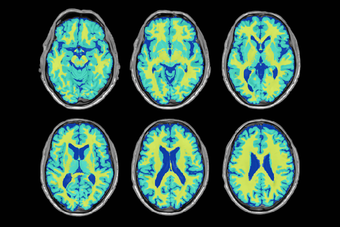
Accurate differentiation of brain tissue types from MR images  is necessary in many neuroscience and clinical applications. Accurate automated tissue segmentation is challenging due to the variability in the tissue intensity profiles caused by differences in scanner models, acquisition protocols, as well as the age of the subjects and presence of pathology. We have developed BISON (Brain tIsue SegmentatiOn pipeliNe), a new pipeline for tissue segmentation using a random forests classifier and a set of intensity and location priors obtained based on T1w images.

BISON Pipelineweb

Execution Example:

python BISON.py -c RF -m Trained_Classifiers/ -o Outputs/ -t Temp_Files/ -e PT -n List.csv -p Trained_Classifiers/ -l 3


Cerebellum and its lobules segmentation

1403365_10151970272361428_660646102_o

The human cerebellum has the highest growth rate of all brain structures during the late fetal and early postnatal life, it plays an integrating role in various neuronal networks, and it shows pathological volume changes in various neuro-psychiatric disorders.

We have developed accurate and fully automatic method to segment cerebellum and it’s lobules using combination of non-linear registration and non-local patch-based label fusion.

References:

  • Katrin Weier, Vladimir Fonov, Karyne Lavoie, Julien Doyon, D Louis Collins. Rapid automatic segmentation of the human cerebellum and its lobules (RASCAL)—Implementation and application of the patch‐based label‐fusion technique with a template library to segment the human cerebellum. Human brain mapping 2014. DOI: 10.1002/hbm.22529
  • Katrin Weier, Vladimir Fonov, Bérengère Aubert-Broche, Douglas L Arnold, Brenda Banwell, D Louis Collins. Impaired growth of the cerebellum in pediatric-onset acquired CNS demyelinating disease. Multiple Sclerosis Journal, 2015 DOI: 10.1177/1352458515615224

Data:

Software:

Brain extraction

beast_picture_crop

Accurate delineation of brain tissue on MRI scan is important step in many image processing pipelines. We have created a method “BEaST: Brain extraction based on nonlocal segmentation technique”, that utilizes library of manually delineated scans for accurate and automatic brain extraction.

References

  1. Eskildsen SF, Coupe P, Fonov V, Manjon JV, Leung KK, Guizard N, Wassef SN, Ostergaard LR, Collins DL; Alzheimer’s Disease Neuroimaging Initiative. BEaST: brain extraction based on nonlocal segmentation technique. Neuroimage. 2012 Feb 1;59(3):2362-73.  DOI: 10.1016/j.neuroimage.2011.09.012

Software

BEaST method is included in the minc-toolkit, available in Software section.

Medial Temporal Lobe (MTL) Segmentation

Our group has developed multiple accurate segmentation methods during years. Our nonlinear patch-based method with error correction is one of the most accurate methods of segmentation of medial temporal lobe structures. Our method reaches 0.901 of Dice similarity with manual tracing for hippocampus which is one of the highest values reported in the literature. Medial temporal lobe atrophy and especially hippocampal atrophy has been one of the most investigated AD biomarkers.

  1. Zandifar, A., Fonov, V. , Coupé, P., Pruessner, J. C.,  & Collins, D. L. (2014). A unified assessment of fully automated hippocampus segmentation methods. Alzheimer’s & Dementia: The Journal of the Alzheimer’s Association10(4), P86.
  2. Zandifar, A., Fonov, V. , Coupé, P., Pruessner, J. C.,  & Collins, D. L. (2015). A quantitative comparison between two manual hippocampal segmentation protocolsAlzheimer’s & Dementia: The Journal of the Alzheimer’s Association, 11(7), P67-P68.
YouTube-logo-full_color