Tissue Classification

Accurate differentiation of brain tissue types from MR images  is necessary in many neuroscience and clinical applications. Accurate automated tissue segmentation is challenging due to the variability in the tissue intensity profiles caused by differences in scanner models, acquisition protocols, as well as the age of the subjects and presence of pathology. We have developed BISON (Brain tIsue SegmentatiOn pipeliNe), a new pipeline for tissue segmentation using a random forests classifier and a set of intensity and location priors obtained based on T1w images.

BISON Pipelineweb

Execution Example:

python BISON.py -c RF -m Trained_Classifiers/ -o Outputs/ -t Temp_Files/ -e PT -n List.csv -p Trained_Classifiers/ -l 3


Brain extraction

beast_picture_crop

Accurate delineation of brain tissue on MRI scan is important step in many image processing pipelines. We have created a method “BEaST: Brain extraction based on nonlocal segmentation technique”, that utilizes library of manually delineated scans for accurate and automatic brain extraction.

References

  1. Eskildsen SF, Coupe P, Fonov V, Manjon JV, Leung KK, Guizard N, Wassef SN, Ostergaard LR, Collins DL; Alzheimer’s Disease Neuroimaging Initiative. BEaST: brain extraction based on nonlocal segmentation technique. Neuroimage. 2012 Feb 1;59(3):2362-73.  DOI: 10.1016/j.neuroimage.2011.09.012

Software

BEaST method is included in the minc-toolkit, available in Software section.

Schizophrenia

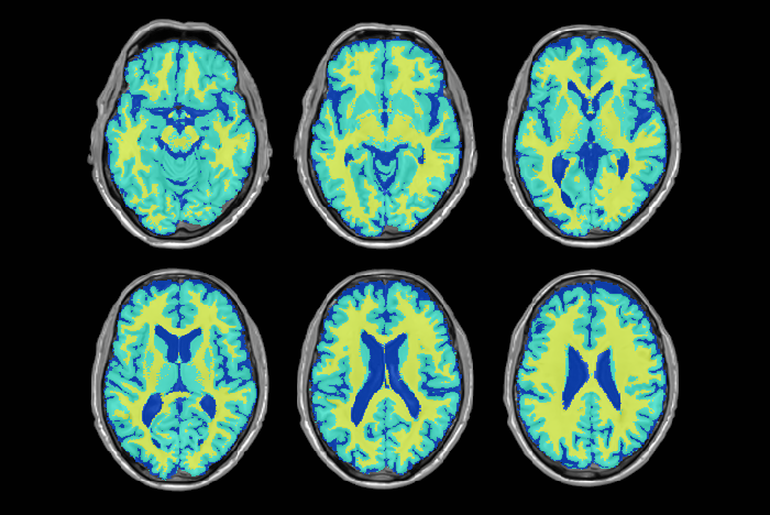
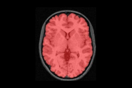
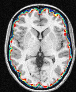
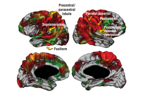
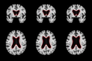
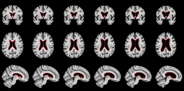
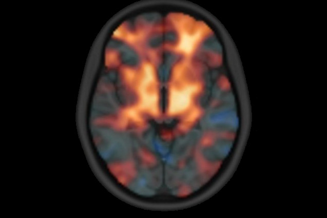
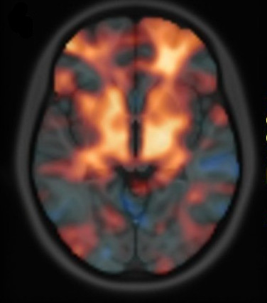
The NIST Lab has collaborated with the Utrecht group headed by Dr. H. Hulshoff Pol for the past number of years [1, 2, 3]. Her group has used my ANIMAL non-linear registration software with voxel based morphometry (VBM) for morphometric analysis of cerebral MRI of patients with schizophrenia. Recently they have provided evidence which suggests that brain structures reflect overlapping and segregating genetic liabilities for schizophrenia and BD. The overlapping smaller white matter volume and common areas of thinner and/or thicker cortex (thinner right orbitofrontal cortex, thinner right (and left) parahippocampus, and thicker temporoparietal and left superior motor cortices) suggest that both disorders share neurodevelopmental roots. They have also demonstrated excessive thinning of the cortex over time, most pronounced in the temporal and frontal areas, with progression across the span of the illness. This excessive thinning appears related to medication and outcome. Their findings were extended to reveal that cannabis use in patients with schizophrenia resulted in additional thinning in the left dorsolateral prefrontal cortex (DLPFC), left anterior cingulate cortex (ACC) and left occipital lobe as compared to those patients with schizophrenia that did not use cannabis after a 5-year follow-up.

In later papers, they have identified focal grey-matter differences in a number of regions when compared to age-matched controls [4]. They have also found focal white matter changes that suggest abnormal inter-hemispheric connectivity of anterior cortical and sub-cortical brain regions, reflecting decreased hemispheric specialisation in schizophrenia [5]. These results have been partially replicated in a twin study [6,7] and a long term follow-up study [8].

References

[1] Hulshoff Pol HE, van Baal GC, Schnack HG, Brans RG, van der Schot AC, Brouwer RM, van Haren NE, Lepage C, Collins DL, Evans AC, Boomsma DI, Nolen W, Kahn RS. Overlapping and segregating structural brain abnormalities in twins with schizophrenia or bipolar disorder. Arch Gen Psychiatry. 2012 Apr;69(4):349–59

[2] van Haren, Neeltje EM, Hugo G. Schnack, Wiepke Cahn, Martijn P. van den Heuvel, Claude Lepage, Louis Collins, Alan C. Evans, Hilleke E. Hulshoff Pol, and René S. Kahn. “Changes in cortical thickness during the course of illness in schizophrenia.” Archives of general psychiatry. 2011; 68(9): 871–880.

[3] M. Rais, N.E.M. van Haren, W Cahn, H.G. Schnack, C. Lepage, D.L. Collins, A. Evans, H. E. Hulshoff Poll and R.S. Kahn, “Cannabis use and progressive cortical thickness loss in areas rich in CB1 receptors during the first five years of schizophrenia,” European Neuropsychopharmacology, 2010 Dec; 20(12): 855–65.

[4] H. E. Hulshoff Pol, H. G. Schnack, R. C. Mandl, N. E. van Haren, H. Koning, D. L. Collins, A. C. Evans, and R. S. Kahn, “Focal gray matter density changes in schizophrenia,” Arch Gen Psychiatry. 2001; 58: 1118–25.

[5] H. E. Hulshoff Pol, H. G. Schnack, R. C. Mandl, W. Cahn, D. L. Collins, A. C. Evans, and R. S. Kahn, “Focal white matter density changes in schizophrenia: reduced inter-hemispheric connectivity,” Neuroimage. 2004; 21: 27–35.

[6] H. Hulshoff Pol, H. G. Schnack, R. C. Mandl, R. G. H. Brans, N. E. van Haren, W. F. C. Baare, C. J. van Oel, D. L. Collins, A. C. Evans, and R. S. Kahn, “Gray and white matter density changes in monozygotic and same-sex dizygotic twins discordant for schizophrenia: a voxel-based morphometry study,” presented at 10th Annual Meeting of the Organization for Human Brain Mapping, Budapest, Hungary, 2004.

[7] H. E. Hulshoff Pol, H. G. Schnack, R. C. Mandl, R. G. Brans, N. E. van Haren, W. F. Baare, C. J. van Oel, D. L. Collins, A. C. Evans, and R. S. Kahn, Gray and white matter density changes in monozygotic and same-sex dizygotic twins discordant for schizophrenia using voxel-based morphometry, Neuroimage. 2006; 31(2): 482–8.

[8] N. E. van Haren, H. E. Hulshoff Pol, H. G. Schnack, W. Cahn, R. C. Mandl, D. L. Collins, A. C. Evans, and R. S. Kahn, “Focal Gray Matter Changes in Schizophrenia across the Course of the Illness: A 5-Year Follow-Up Study,” Neuropsychopharmacology. 2007; 32: 2057–66.

Normal Aging

adni_model_4d_v2

Over the past several years we have collaborated with several individuals to study normal aging. Using Alzheimer’s disease, amnesic mild cognitive impairment and healthy individuals we have demonstrated that interhemispheric coupling may be regarded as a flexible mechanism that can improve the brain’s ability to meet processing demands for high cognitive demand in normal aging and for low cognitive demand in AD [1].

In a recent study the links between cognitive ability and cortical tissue volume in old age were investigated. Evidence from this research warns against an exclusive reliance on the causal link between cognitive function and the cortical tissue in old age based on assumptions of the aging process. Preservation of cortical tissue thickness in old age is not a foundation for successful cognitive aging, but rather reflects a lifelong association [2].

Reference

[1] J. Ansado, D.L. Collins, S. Joubert, V.S. Fonov, O. Monchi, S.M. Brambati, F. Tomaiuolo, M. Petrides, S. Faure, Y. Joanette, Yves. “Interhemispheric coupling improves the brain’s ability to perform low cognitive demand tasks in Alzheimer’s disease and high cognitive demand tasks in normal aging”, Neuropsychology, Vol 27(4), Jul 2013, 464–480.

[2] Karama S, Bastin ME, Murray C, Royle NA, Penke L, Muñoz Maniega S, Gow AJ, Corley J, Valdés Hernández Mdel C, Lewis JD, Rousseau MÉ, Lepage C, Fonov V, Collins DL, Booth T, Rioux P, Sherif T, Adalat R, Starr JM, Evans AC, Wardlaw JM, Deary IJ. Childhood cognitive ability accounts for associations between cognitive ability and brain cortical thickness in old age. Mol Psychiatry. 2014 May;19(5):555–9.

Epilepsy

In some patients with refractory epilepsy that are candidates for surgery, intracranial EEG is recorded to precisely localize the epileptic focus. To record intracranial EEG, multiple depth electrodes are surgically implanted through holes in the skull, each with 8–15 equally spaced contacts. Current implantation planning consists of visual inspection of the patient’s MRI & CTA, visually searching for paths to the targets while avoiding vessels. This procedure is sub-optimal since estimation of the number of electrodes needed to sample a region and the precise location of each contact, which is paramount to accurately identify the focus and its extent, cannot be considered.

The goal of this project, conducted by Dr. Zelmann, one of my postdoctoral fellows, is to evaluate the clinical use of optimized depth electrode planning. We hypothesize that the use of our computed aided procedure will increase the accuracy and amount of information obtained during EEG intracranial investigation while constraining the trajectories to safe paths.

We have developed a procedure to optimize electrode location that would enable us to obtain complete coverage of a lesion or region of interest and surrounding neocortical grey matter, while minimizing the risk of approaching vessels and other critical structures. To this end, we automatically segment MRI, CT and CTA data; we model each electrode as a cylinder to assess risk; we estimate the contribution of individual contacts to record EEG; we compute an aggregated score for each electrode and a global score, obtaining the best cohort as the combined set of electrodes that remain at a safe distance.

To allow its clinical use during planning and surgical implantation, the procedure is integrated into an image-guided neuronavigation system developed in our research laboratory over the past decade, which is called IBIS (Interactive Brain Imaging System; Mercier et al., Int J CARS. 2011;6(4):507–522). IBIS allows planning and navigation based on preoperative medical image data. The best cohort and a list of possible electrodes per target (ordered in terms of risk and recorded EEG) is generated and displayed in IBIS and will be available during planning and in the OR. The surgeon can visually review trajectories, decide between one of the automatic trajectories and the manual one and, if necessary, re-plan a trajectory in almost real time.

Preliminary results [1] presented at an international workshop on clinical image-based procedures, suggest that automatic planning allows recording from a larger volume than manually planned trajectories (p<0.01) and from more temporal grey matter (p<0.001), while remaining further away from segmented vessels (p<0.01). We are currently evaluating the procedure in retrospective clinically acquired imaging data from 20 patients with electrodes implanted in MTL regions. We are comparing visual and automatic trajectories by estimating volume recording from the target volumes (in this case: amygdala and hippocampus) and neocortical temporal grey matter as well as distance to vessels and other critical structures.

Reference

[1] Zelmann, R., Beriault, S., Mok, K., Haegelen, C., Hall, J., Pike, G. B., Olivier, A & Collins, D. L. (2014). Automatic Optimization of Depth Electrode Trajectory Planning. In Clinical Image-Based Procedures. Translational Research in Medical Imaging (pp. 99–107). Springer International Publishing.

[2] R. Zelmann, S. Beriault, K. Mok, C. Haegelen, J. Hall, G.B. Pike, A. Olivier & D.L. Collins, “Improving Recorded Volume in Mesial Temporal Lobe by Optimizing Stereotactic Intracranial Electrode Implantation Planning” submitted to International Journal of Computer Assisted Radiology and Surgery (CARS-D-14–00251), Oct 9, 2014.

[3] Zelmann, R., Beriault, S., Mok, K., Haegelen, C., Hall, J., Pike, G. B., Olivier, A & Collins, D. L. (2014). Automatic Optimization of Depth Electrode Trajectory Planning. In Clinical Image-Based Procedures. Translational Research in Medical Imaging (pp. 99–107). Springer International Publishing.

[4] R. Zelman, D.L. Collins, “Automatic Optimization of Depth Electrode Trajectory Planning”, MICCAI 2013 Workshop on Clinical Image-based Procedures: Translational Research in Medical Imaging

[5] C. Haegelen, P. Perucca, C.-E. Châtillon, L. Andrade-Valença, R. Zelmann, J. Jacobs, D. L. Collins, F. Dubeau, A. Olivier and J. Gotman. “High-frequency oscillations, extent of surgical resection and surgical outcome in drug-resistant focal epilepsy”, Epilepsia 2013 May;54(5):848–57.

[6] Zelmann, R., S. Beriault, M. M. Marinho, K. Mok, J. A. Hall, N. Guizard, C. Haegelen, A. Olivier, G. B. Pike, and D. L. Collins. “Improving recorded volume in mesial temporal lobe by optimizing stereotactic intracranial electrode implantation planning.” International journal of computer assisted radiology and surgery (2015): 1–17.

Parkinson’s Disease

Parkinson’s disease (PD) is the second most common neurodegenerative disease, after Alzheimer’s disease, worldwide. At the NIST Lab, we are dedicated to explore potential structural biomarkers for the disease, as well as proposing image processing techniques for the surgical treatment of PD.


Projects include:

Stereotaxic surgery for movement disorders

Autism Spectrum Disorder

Autism Spectrum Disorder (ASD) is a set of neurological disorders characterized by impaired social communication and interaction, as well as restricted, repetitive patterns of behaviour, interests and activities. It is commonly diagnosed at around the ages of 3 to 4 years, developing tools that could aid in obtaining a diagnosis as early as 6 or 12 months of age would allow an early therapy that could potentially increase the adaptation to society of children with ASD.

At the NIST, we have utilized voxel-wise image processing methods to investigate the brain development of children at high-risk of ASD during the first 2 years of life. This work has led to observations of significant differences in the growth trajectories of several regions in the brain between children who are diagnosed with ASD at 2 years of age and normal controls.

HIV Neurodegeneration

HIV enters the brain soon after seroconversion and potentially causes cognitive impairment. Although the incidence of severe dementia has been reduced, perhaps due to effective HIV treatment, the prevalence of mild to moderate cognitive impairment appears to be increasing. It has been reported that 30-50% of HIV+ patients with well-controlled infections show cognitive deficits. Several factors are thought to contribute to this brain injury. However, the literature has yet to produce a clear consensus of the mechanisms that may underlie brain injury.

HIV
Brain volume loss associated with a history of severe HIV-related immunosuppression.

At NIST, we have utilized novel neuroimaging methods with complementary strengths, deformation-based morphometry, voxel-based morphometry and cortical modeling, to investigate the effects HIV has on brain structure and function. Here, we observed regionally specific patterns of reduced cortical and subcortical volumes in the HIV+ group. White matter loss and subcortical atrophy was related a history of more severe immunosuppression, while cortical thickness reductions were related to poorer neuropsychological test performance. The findings suggest that distinct mechanisms may underlie cortical and subcortical injury, and argues for the potential importance of early HIV treatment in protecting long term brain health.

Abstracts and Conference Presentations:

R. Sanford, A.L. Fernandez Cruz, L.K. Fellows, B.M. Ances, D.L. Collins, Regionally Specific Cortical Thinning in HIV+ Patients in the cART Era, 2016 Conference on Retroviruses and Opportunistic Infections (CROI), February 2016, Boston, Massachusetts (pdf)

Alzheimer’s

Alzheimer’s disease (AD) is a progressive neurodegenerative disease, which is the most common cause of dementia. Its prevalence for people aged 65-70 y is 1%, while it is 7% for the 75-84 y group, and 26% among those aged 90 and older. In Canada, persons over age 65 will make up to 15% of the population by 2016, but this amount is estimated to reach 23% by 2041. It is assumed that the prevalence of AD will quadruple by 2050 resulting in great financial burden.

It is believed that the pathophysiological process of AD begins well before the diagnosis of the dementia. Like many other neurodegenerative diseases, early treatment, before occurrence of too much irreversible degeneration of brain tissue, can be more effective. However, early diagnosis of Alzheimer’s disease is currently almost impossible. The well-cited biomarker model that the structural MRI begins showing abnormality at the preclinical stage and rises significantly in MCI stage, which makes it an interesting candidate for prognosis of dementia onset. Our group with sophisticated image analysis techniques followed by statistical analysis tries to make the diagnosis and prediction possible at the level of an individual person.

Projects include:

Segmentation of White Matter Hyperintensities

Medial Temporal Lobe (MTL) Segmentation

YouTube-logo-full_color