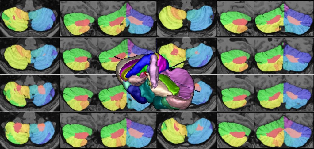
The human cerebellum has the highest growth rate of all brain structures during the late fetal and early postnatal life, it plays an integrating role in various neuronal networks, and it shows pathological volume changes in various neuro-psychiatric disorders.
We have developed accurate and fully automatic method to segment cerebellum and it’s lobules using combination of non-linear registration and non-local patch-based label fusion.
References:
- Katrin Weier, Vladimir Fonov, Karyne Lavoie, Julien Doyon, D Louis Collins. Rapid automatic segmentation of the human cerebellum and its lobules (RASCAL)—Implementation and application of the patch‐based label‐fusion technique with a template library to segment the human cerebellum. Human brain mapping 2014. DOI: 10.1002/hbm.22529
- Katrin Weier, Vladimir Fonov, Bérengère Aubert-Broche, Douglas L Arnold, Brenda Banwell, D Louis Collins. Impaired growth of the cerebellum in pediatric-onset acquired CNS demyelinating disease. Multiple Sclerosis Journal, 2015 DOI: 10.1177/1352458515615224
Data:
- Preprocessed MRI scans and segmentation labels in MINC2 format are available for download: rascal_raw_library_20170817.zip 1.7Gb
- Library in the format suitable for use with the patch-based fusion segmentation pipeline, is also available for download: rascal_t1w_seg_library_20170817.zip 1.6 Gb
Software:
-
All the software used in cerebellum segmentation method is included on minc-toolkit v2, available in Software Section.
patch-based fusion segmentation pipeline, can be used to segment pre-processed MRI scans.

