Colin 27 Average Brain, Stereotaxic Registration Model, high-resolution version 2008
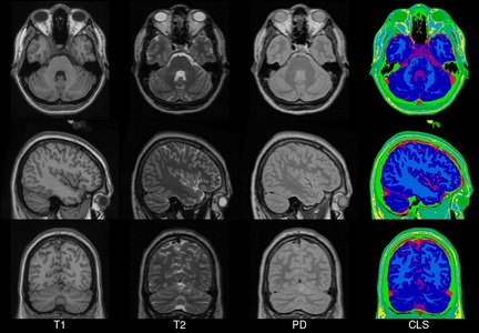
The anatomical phantom is derived from T1, T2, PD-weighted images formed from the average of 27, 11 and 12 scans respectively, of the same normal subject. These volumes are defined at a 0.5mm isotropic voxel grid in Talairach space, with dimensions 362*434*362 (XxYxZ) and start coordinates −90,−126,−72 (x,y,z). A discrete phantom was created by storing the label of the most important fraction class at each voxel location
Publications
- Holmes CJ, Hoge R, Collins DL, Woods R, Toga AW, Evans AC. “Enhancement of MR images using registration for signal averaging.” J Comput Assist Tomogr. 1998 Mar-Apr;22(2):324–33. https://dx.doi.org/10.1097/00004728-199803000-00032
- B Aubert-Broche, AC Evans, and DL Collins, “A new improved version of the realistic digital brain phantom,” NeuroImage, vol. 32, no. 1, pp. 138–45, 2006. https://www.ncbi.nlm.nih.gov/pubmed/16750398
License
Copyright (C) 1993–2009 Louis Collins, McConnell Brain Imaging Centre, Montreal Neurological Institute, McGill University. Permission to use, copy, modify, and distribute this software and its documentation for any purpose and without fee is hereby granted, provided that the above copyright notice appear in all copies. The authors and McGill University make no representations about the suitability of this software for any purpose. It is provided “as is” without express or implied warranty. The authors are not responsible for any data loss, equipment damage, property loss, or injury to subjects or patients resulting from the use or misuse of this software package.
Download
Download archives containing average t1w, t2w and pdw scan, and discrete tissue classification: 1: Cerebro-spinal fluid, 2: Gray Matter, 3: White Matter, 4: Fat, 5: Muscles, 6: Skin and Muscles, 7: Skull, 9: Fat 2, 10: Dura, 11: Marrow, 12: Vessels
MINC1 222MB MINC2 223MB NIFTI 291MB
Fuzzy segmentation
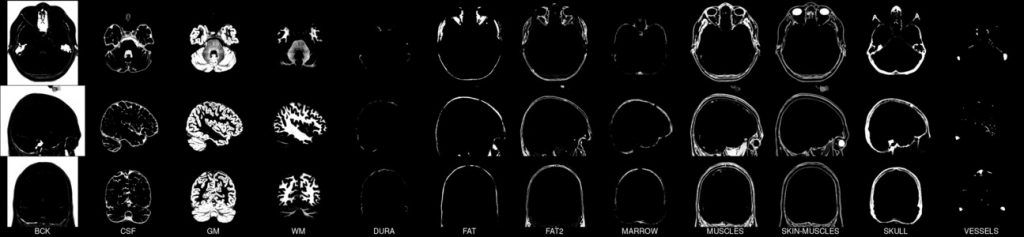
Download archives containing 12 volumetric fuzzy volumes that define the spatial distribution for different tissues where voxel intensity is proportional to the fraction of tissue within the voxel (the integral of all tissue components is equal to 1).
MINC1 54MB MINC2 57MB NIFTI 90MB