MNI Average Brain (305 MRI) Stereotaxic Registration Model
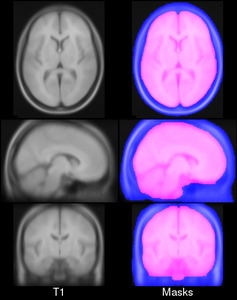 This is a version of the MNI Average Brain (an average of 305 T1-weighted MRI scans, linearly transformed to Talairach space) specially adapted for use with the MNI Linear Registration Package (mni_reg).
Methods
In order to overcome the idiosyncrasies of using a single subject brain as a template, in the early 1990s Evans and colleagues introduced the concept of a statistical MRI atlas for brain mapping (Evans et al., 1992a,b, 1993). The MNI305 atlas was constructed in two steps.
First, anatomical landmarks were manually identified in T1-weighted MRI scans from young healthy subjects (239 males, 66 females, age 23.4 +/- 4.1 years). These landmarks were chosen from the Talairach and Tournoux atlas and thus the final aver- age and space approximated Talairach space. Landmarks from each subject were fitted together via least-squares linear regression that matched the resulting AC-PC line to the original Talairach and Tournoux atlas. This yielded a first-pass average T1-weighted MRI volume.
Second, each native MRI volume was automatically mapped to the manually-derived average MRI to reduce the impact of order effects, manual errors and to create a sharper average. The mapping was not performed according to Talairach’s piecewise linear model but used a whole-brain linear (9-parameter) image similarity residual (Collins et al., 1994). The resultant template is thus an approx- imation of the original Talairach space and the Z-coordinate is approximately +3.5 mm relative to the Talairach coordinate. This process resulted in the original MNI305 atlas that has sub- sequently defined the MNI space. Note that, under constraints of linear alignment, residual non-linear anatomical variability across subjects gives rise to a “virtual convolution” (Evans et al., 1993) that somewhat enlarges the template compared with most individual brains.
Publications
- Collins, D.L., Neelin, P., Peters, T.M., Evans, A.C., 1994. Automatic 3-D intersubject reg- istration of MR volumetric data in standardized Talairach space. J. Comput. Assist. Tomogr. 18 (2), 192–205.
- Evans, A.C., Collins, D.L., Milner, B., 1992a. An MRI-based stereotaxic atlas from 250 young normal subjects. Proc 22nd Annual Symposium, Society for Neuroscience, 18, p. 408.
- Evans, A.C., Marrett, S., Neelin, P., Collins, D.L., Worsley, K., Dai, W., Milot, S., Meyer, E., Bub, D., 1992b. Anatomical mapping of functional activation in stereotactic coordi- nate space. NeuroImage 1 (1), 43–63.
- Evans, A.C., Collins, D.L., Mills, S.R., Brown, E.D., Kelly, R.L., Peters, T.M., 1993. 3D statis- tical neuroanatomical models from 305 MRI volumes. Proc IEEE-Nuclear Science Symposium and Medical Imaging Conference, pp. 1813–1817.
License
Copyright (C) 1993–2009 Louis Collins, McConnell Brain Imaging Centre, Montreal Neurological Institute, McGill University. Permission to use, copy, modify, and distribute this software and its documentation for any purpose and without fee is hereby granted, provided that the above copyright notice appear in all copies. The authors and McGill University make no representations about the suitability of this software for any purpose. It is provided “as is” without express or implied warranty. The authors are not responsible for any data loss, equipment damage, property loss, or injury to subjects or patients resulting from the use or misuse of this software package.
Download
Download archives containing average t1w model, brain mask and head mask: MINC1 3.6MB MINC2 3.7MB NIFTI 4.8MB